Round bacteria that grow in clusters are called staphylococci. More specifically, round bacteria are called cocci (singular: coccus), and when cocci group together in irregular, grape-like clusters, that arrangement is the defining visual signature of the genus Staphylococcus. If you've seen this term on a lab report or in a microbiology textbook and wondered what it actually means, here's a clear breakdown of the terminology, what causes that clustering, and why it matters in practical food safety.
Round Bacteria in Clusters Are Called Cocci and Staphylococci

Marcus Reeves
1 Jul 2026
The microbiology terminology for cocci
In bacterial morphology, shape is the first thing you classify. The three basic shapes are cocci (round), bacilli (rod-shaped), and spirilla (spiral). Cocci are simply spherical or ovoid bacteria. The word comes from the Greek kokkos, meaning grain or berry, which gives you a good mental image of what they look like under a microscope.
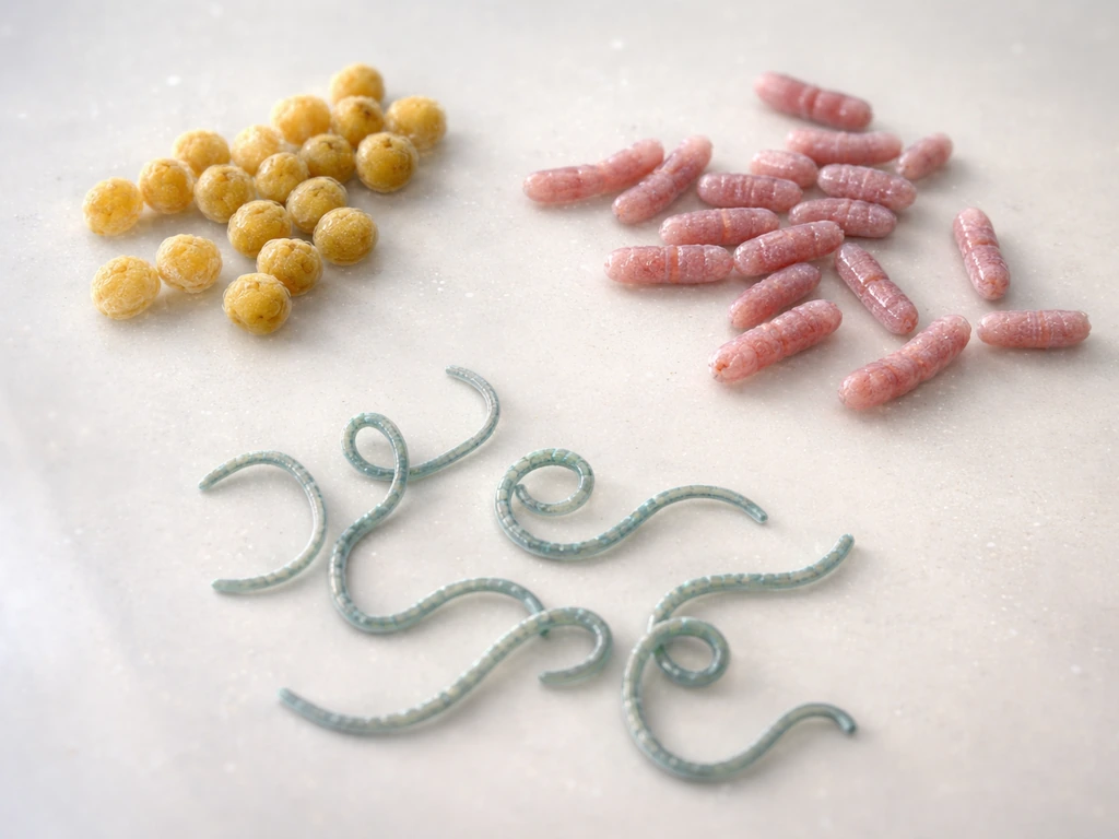
Beyond shape, cocci are also classified by how they arrange themselves after dividing. This is where the terminology gets more precise. The main arrangements you'll encounter are:
- Diplococci: cocci in pairs
- Streptococci: cocci in chains
- Tetrads: groups of four cocci arranged in the same plane (associated with genera like Micrococcus)
- Sarcinae: groups of eight cocci arranged into a cube-like packet
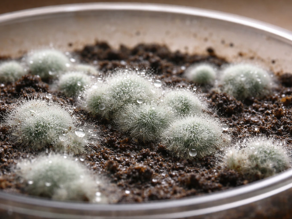
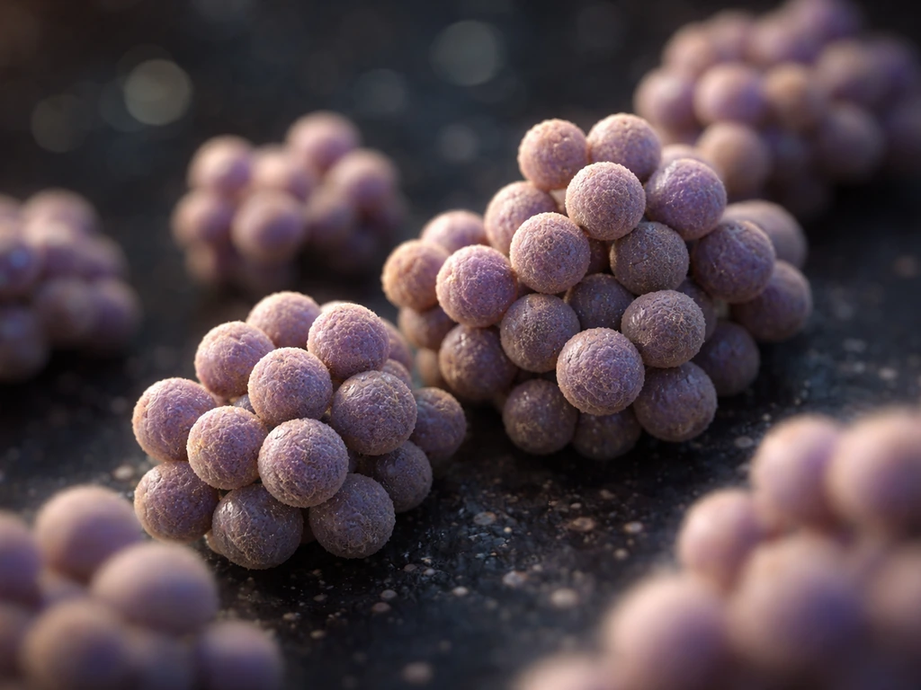
- Staphylococci: cocci in irregular, grape-like clusters
Each of these arrangement terms describes both a visual pattern and, in some cases, an entire genus. When someone says 'staphylococci,' they're using a term that's simultaneously a morphological description (clustered cocci) and a reference to the Staphylococcus genus.
Why bacteria form clusters instead of chains
Clustering isn't random. It comes down to how cells divide and whether they stay attached afterward. When a coccus divides, the two daughter cells can either separate cleanly, stay connected in one plane (forming a chain), or remain attached across multiple planes. Staphylococci divide in multiple planes and the daughter cells stay partially attached, which produces that characteristic irregular, grape-like pile of cells.
Streptococci, by contrast, divide in a single plane and cells stay connected end-to-end, which is exactly why they form chains rather than clusters. This distinction is genuinely useful because it narrows down what genus you might be dealing with when you look at a slide. Chains point toward streptococci; irregular clusters point toward staphylococci.
In practice, you'll sometimes see staphylococci appearing in pairs or even short chains on a Gram stain, especially if they were grown in liquid media or if the smear is dense. The grape-like cluster is the hallmark, but don't expect every field of view to look like a perfect textbook illustration.
How to confirm the morphology in a lab
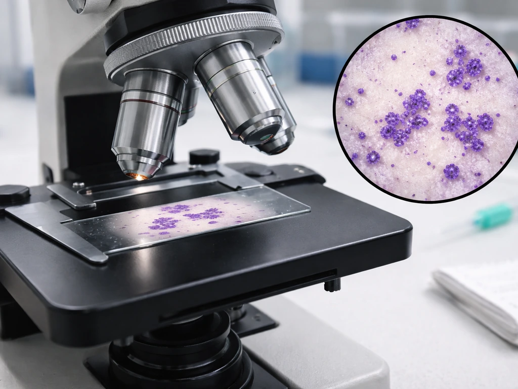
If you're trying to identify an organism based on morphology, the Gram stain is your starting point, not your finish line. A Gram stain tells you two things immediately: whether the bacterium is Gram-positive (purple) or Gram-negative (pink/red), and the shape and arrangement of the cells. Staphylococci are Gram-positive cocci in clusters, so on a properly prepared smear you'd see purple, round cells in irregular groupings.
The CDC uses a morphology-first workflow for presumptive bacterial identification: you start with the Gram reaction, then look at the cell shape, then assess the arrangement (pairs, chains, tetrads, or clusters). Clusters of Gram-positive cocci push you toward staphylococci in that decision tree.
That said, morphology alone isn't enough for a confirmed ID. You need to combine Gram stain results with other characteristics like colonial morphology on plates, hemolysis patterns on blood agar, and biochemical tests (such as the catalase test and the coagulase test, which is the key test for distinguishing Staphylococcus aureus from other staphylococci). Gram stain arrangement is the first filter, not the final answer.
Common cluster-forming cocci groups
The most clinically and food-safety-relevant cluster-forming cocci belong to the genus Staphylococcus. Within that genus, there are two broad groups you'll commonly encounter:
Staphylococcus aureus
S. aureus is the most well-known member. It's a Gram-positive, facultative anaerobe (meaning it can grow with or without oxygen, though it prefers oxygen-rich conditions). It's the primary culprit in staphylococcal food poisoning and produces heat-stable enterotoxins that survive normal cooking temperatures. Its classic appearance is Gram-positive cocci in grape-like clusters.
Coagulase-negative staphylococci (CoNS)
This group includes species like Staphylococcus epidermidis. They also appear as Gram-positive cocci in clusters (sometimes pairs or tetrads), but they test negative for coagulase, which is the biochemical feature that separates them from S. aureus. CoNS are less associated with food poisoning but are relevant in clinical and environmental microbiology contexts.
Staphylococci are also sometimes confused with micrococci, which are Gram-positive cocci that tend to form tetrads and are generally considered non-pathogenic. The arrangement difference (tetrads vs. irregular clusters) is one visual clue, but again, biochemical testing is needed to confirm.
How environmental conditions affect cluster-forming bacteria
Understanding the morphology of staphylococci is more useful when you pair it with knowledge of the conditions that let them grow. S. aureus in particular is remarkably tolerant of challenging environments, which makes it a significant concern in food settings.
| Condition | S. aureus Growth Range | Practical Notes |
|---|---|---|
| Temperature | 7–48°C (45–118°F) | Fastest growth and toxin production at 20–37°C (68–99°F); standard refrigeration below 4.4°C slows growth significantly |
| pH | Approximately 4.5–9.3 | Grows across a wide pH range; acidic conditions closer to 4.5 inhibit but don't eliminate growth |
| Water activity (a_w) | As low as 0.83 for growth; higher required for toxin production | More tolerant of low moisture than many other food pathogens; survives in cured and dried foods |
| Oxygen | Facultative anaerobe | Grows with or without oxygen; prefers aerobic conditions but can survive in vacuum-packed or modified-atmosphere environments |
The wide temperature and water activity tolerances are what make S. aureus particularly tricky in food environments. It can grow in conditions that would stop other pathogens, and it can persist in relatively dry foods where you might not expect bacterial growth. Penicillin is different from staphylococci because it grows naturally in specific molds, primarily Penicillium species found in soil and decaying plant material where does penicillin grow naturally.
The most dangerous window is the food safety danger zone: 40–140°F (4.4–60°C). Within that range, and especially between 68–99°F (20–37°C), staphylococci multiply rapidly and produce enterotoxins. If food sits at room temperature long enough, the toxin can reach harmful levels even if you later reheat or cook the food, because the toxins themselves are heat-stable and not destroyed by normal cooking.
Practical relevance to food safety and contamination prevention
Knowing that cluster-forming cocci like staphylococci are the culprit behind a significant category of foodborne illness changes how you think about food handling. The toxin is preformed in the food before you eat it, the food may look and smell completely normal, and onset of symptoms is fast: typically 1–6 hours after eating contaminated food. There's no warning sign you can rely on.
The most important control point is time and temperature. USDA FSIS refrigeration guidance explains that keeping food at safe temperatures slows bacterial growth, helping limit the risk from bacteria that could contaminate food blank" rel="noopener noreferrer">The most important control point is time and temperature.. Staphylococci can colonize food via skin contact (they're common on human skin and nasal passages), and if contaminated food is left in the danger zone, they multiply and produce toxin. The practical rules:
- Refrigerate cooked and prepared foods promptly, below 40°F (4.4°C). Don't leave food sitting at room temperature for more than two hours.
- Keep hot foods hot (above 140°F / 60°C) and cold foods cold. The danger zone is where staph multiplies fast.
- Don't assume reheating will fix it. If toxin has already formed, cooking won't destroy it. The food needs to be discarded.
- Wash hands thoroughly before handling food, especially after touching your face or skin. Staphylococci are common on skin and can transfer directly to food.
- Sanitize food-contact surfaces regularly. A bleach solution of about 1 tablespoon of chlorine bleach per gallon of hot water is a standard approach for surface sanitizing.
- Avoid mixing freshly cooked hot foods with cold foods without ensuring the entire mixture reaches a safe serving or storage temperature.
It's also worth connecting the morphology to the bigger picture: staphylococci are pus-forming bacteria, and the cluster-forming arrangement (grape-like cocci) is part of how microbiologists identify them quickly in clinical and food lab settings. If you're working in food service or food production and you see a lab report referencing 'Gram-positive cocci in clusters,' that's a signal to take the contamination source seriously and trace it back to handling practices or environmental conditions.
Biofilm formation is another consideration in food processing environments. Staphylococci can form biofilms on food-contact surfaces, making them harder to remove with standard cleaning. In general, biofilms grow wherever bacteria can attach to moist surfaces, including food-contact areas and medical equipment where do biofilms grow. This is one reason sanitizing protocols go beyond simple soap-and-water cleaning, especially in facilities handling ready-to-eat foods.
The bottom line: when you know that round bacteria in clusters are called staphylococci, and you understand the conditions that let them thrive (warm temperatures, moderate moisture, and time), you have the foundation to make smarter storage and handling decisions. The terminology isn't just academic. It maps directly onto which bacteria you're dealing with, how they behave, and what actually stops them.
FAQ
If I see round purple cocci in clusters on a Gram stain, is that automatically staphylococci?
Not necessarily. If the smear is too thick, bacteria can clump artificially, making cocci look clustered even when they are not arranged that way naturally. Use an appropriately thin smear and confirm the arrangement across multiple microscope fields before calling it “clusters.”
Why might staphylococci look like pairs or short chains instead of perfect grape-like clusters?
Yes, but the “typical” grape-like look can be subtle. If cells are partially damaged, the fixation is too harsh, or incubation conditions differ, clusters may appear as irregular pairs, small packets, or occasional short chains. That is why labs treat arrangement as a prescreening clue, not a final ID.
How can I tell staphylococci apart from micrococci when both are Gram-positive cocci?
Micrococci can be confusing because they are also Gram-positive cocci. A key visual distinction is tetrad formation (micrococci) versus multi-plane irregular clusters (staphylococci), but since overlap happens, you still need confirmatory testing such as catalase and coagulase (and additional species tests if required).
What confirmatory tests should I consider after seeing Gram-positive cocci in clusters?
Gram positivity alone is not enough, because other Gram-positive cocci can look similar in shape. The practical workflow is, Gram reaction first, then arrangement, then confirm with simple tests like catalase (staphylococci are catalase-positive) before moving to species-level assays.
Which tests are most useful for distinguishing S. aureus from other clustered Gram-positive cocci?
For suspect staphylococcal isolates, catalase and coagulase results guide the next steps. Catalase helps separate staphylococci from catalase-negative cocci, and coagulase helps distinguish S. aureus from many coagulase-negative staphylococci (CoNS).
Can staphylococci findings be misleading if my sample contains a mixed culture?
Yes. In real food samples, you may detect Gram-positive cocci in clusters from multiple organisms, or you may get mixed morphology if the sample was contaminated by more than one source. Mixed cultures can produce “mixed” patterns, so take colonies from isolation plates and test them individually.
Do growth conditions affect how staphylococci clusters appear on a smear?
Incubation temperature and growth conditions can change how tightly cells pack after division, so the exact cluster size and appearance can vary. If you only have a single growth condition, consider repeating or checking colonies grown under standard conditions used for the diagnostic workflow.
How does biofilm formation affect lab identification and food-safety decisions?
Biofilm presence changes cleaning effectiveness, but it does not change the basic morphology rules. You can still see Gram-positive cocci, yet routine cleaning may leave cells behind, allowing regrowth. This is why facilities often verify sanitation by environmental sampling, not just by assuming a surface is clean after washing.
If bacteria are killed by cooking, why can staphylococcal food poisoning still occur?
The toxin issue is time-dependent rather than smell- or appearance-dependent. Even if later reheating kills the bacteria, preformed staphylococcal enterotoxins can remain active because they are heat-stable. The best prevention is preventing time in the temperature danger zone and preventing skin-related contamination.
What should I check operationally if a report suggests staphylococcal contamination?
If suspected, treat it as a time-temperature control failure. Document how long the food sat, what temperatures were reached, and whether holding equipment was functioning properly. Then trace potential contamination routes such as hand contact, nasal shedding during prep, and cross-contact from food-contact surfaces.
Next Article
Where Does Penicillin Grow Naturally? Habitat and Conditions
Explains where penicillin-producing Penicillium grows naturally and the damp, nutrient, oxygen conditions that enable co