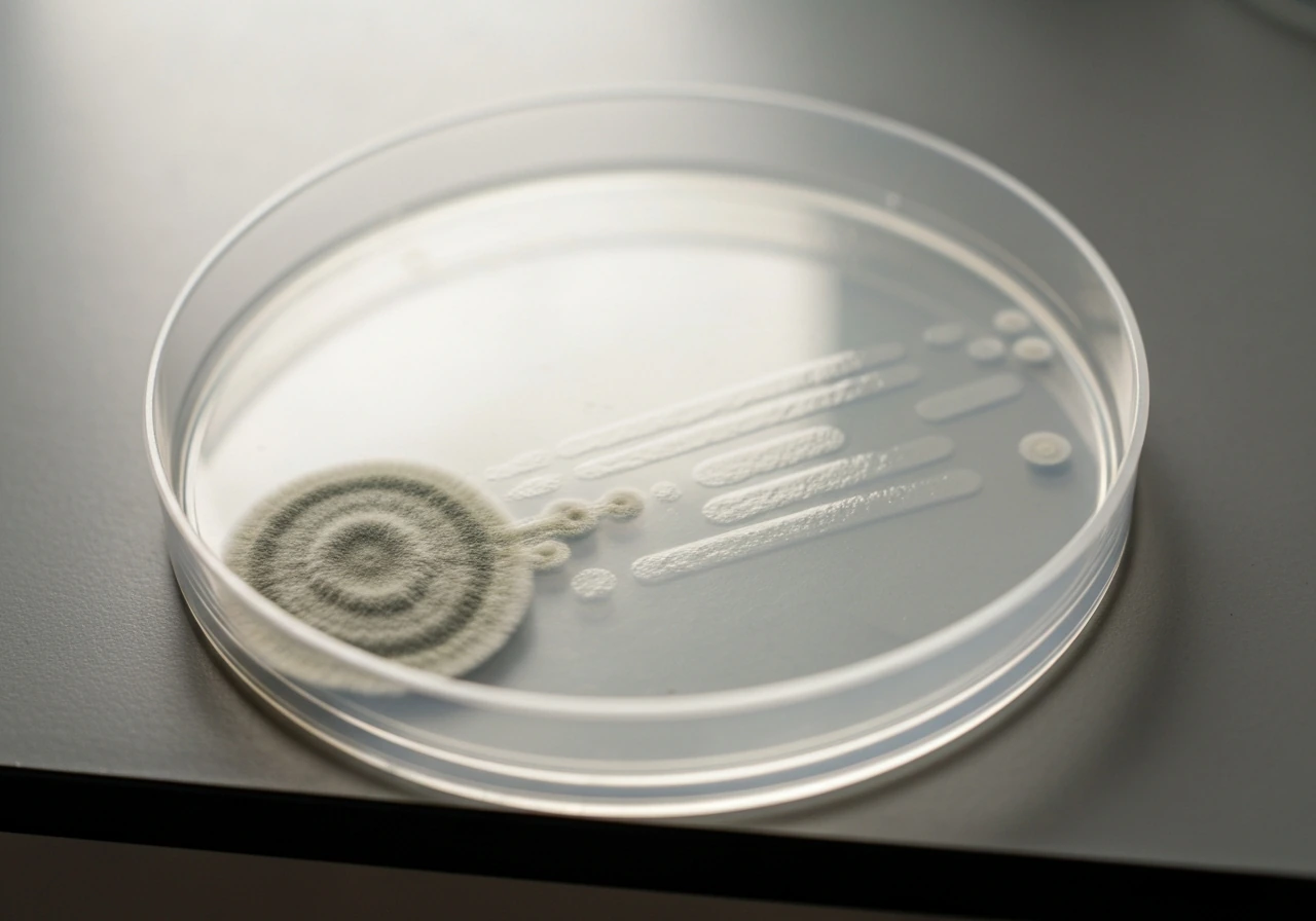
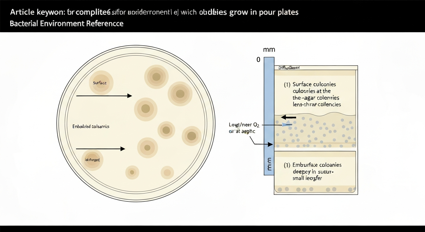
In a pour plate, colonies grow in three distinct locations: on the surface of the solidified agar, just below the surface in the upper agar layer, and fully embedded within the agar matrix. Surface colonies are larger and circular because they have direct access to atmospheric oxygen. Subsurface and embedded colonies are smaller and lens-shaped (lenticular) because they are physically trapped in the gel, cut off from free oxygen, and dependent on diffusion for both nutrients and gas exchange. All three populations count toward your total viable count, and understanding why each forms where it does is the key to interpreting your plates correctly.
Where Do Colonies Grow in Pour Plates: Surface, Subsurface

Marcus Reeves
16 Jul 2026
What a pour plate actually is
A pour plate is made by adding a measured volume of inoculum (typically 1 mL) directly to a sterile, empty petri dish, then pouring 12 to 15 mL of molten agar cooled to 45 ± 1 °C on top. The two are mixed immediately by alternating rotation and back-and-forth motion, then the plate is set on a level surface to solidify. Once set, the plate is inverted and incubated. The inversion step prevents condensation from dripping onto the surface and distorting colonies.
The critical feature of this method is that the inoculum is distributed throughout the agar volume during mixing, not just spread across the top. This means organisms end up at many different depths, and colony position after incubation reflects a combination of where cells were during gelation and what oxygen or nutrient conditions they experienced once the plate solidified. That is why the same sample can simultaneously produce large, spreading surface colonies and tiny, lens-shaped colonies buried deep in the agar.
The three colony locations defined
Every pour plate you read will show colonies in one or more of these three positions. Knowing the vocabulary and visual markers for each prevents misidentification and miscounting.
- Surface colonies: growing at the agar–air interface, typically the largest and most morphologically distinct colonies on the plate.
- Subsurface colonies: growing just below the top of the agar, often appearing as slightly flattened or lenticular colonies visible through the gel.
- Embedded colonies: fully enclosed within the agar matrix, usually small, lens-shaped, and sometimes only visible under a plate magnifier or with low-angle lighting.
The distinction matters practically. If you only count the obvious surface colonies, you will underestimate your viable count. Standards including the FDA Bacteriological Analytical Manual (BAM) Chapter 3 are explicit that both surface and embedded colonies must be counted toward the total, within the acceptable range of 15 to 300 CFU per plate. For regulatory context and links to APHA, AOAC, ISO and USDA methods used for aerobic plate count and pour‑plate enumeration, see the BAM / FDA landing page, references to APHA, AOAC, ISO and USDA methods used for APC/pour‑plate enumeration BAM / FDA landing page — references to APHA, AOAC, ISO and USDA methods used for APC/pour‑plate enumeration.
Surface colonies: biggest, boldest, oxygen-fed
Surface colonies form at the air–agar interface, which means they have unrestricted access to atmospheric oxygen throughout incubation. This is why they grow larger, produce more characteristic pigmentation, and show the full morphological features (raised form, entire margins, surface texture) that you rely on for identification. In most standard count plates, these are the colonies you will spot first.
Cells that end up at the surface are those that were near the top of the agar when gelation occurred, or in some cases cells that were deposited directly at the air–agar interface before the agar fully solidified. Because gelation happens quickly after pouring (agar sets at approximately 42 °C), cells do not have time to settle significantly if mixing and pouring are done correctly according to BAM instructions.
Typical organisms that produce prominent surface colonies in pour plates include Staphylococcus aureus, members of the Enterobacteriaceae family, Pseudomonas species, and most facultative anaerobes that grow robustly under aerobic conditions. Fast-growing aerobes and surface spreaders like certain Bacillus species can produce very large surface colonies that merge with neighbors, a problem covered in the troubleshooting section below.
Subsurface colonies: the lens-shaped layer just below
Subsurface colonies form in the zone immediately below the agar surface, usually within the first millimeter or so of gel depth. They are the most commonly overlooked colony type in student and early-career lab work. Visually, they appear lenticular (lens- or biconvex-shaped) when viewed from above or through the side of the plate, because the agar constrains their expansion into a flat disc and redirects growth laterally into an oblate form.
These colonies represent cells that were trapped just below the surface during solidification. They have some access to diffused oxygen but less than surface colonies. Microelectrode and diffusion modeling data show that oxygen in agar is depleted to near-anoxic conditions within roughly 500 µm of depth when microorganisms are actively respiring, so subsurface colonies are growing in a progressively oxygen-limited microenvironment compared to their surface counterparts. de Beer D., Glud A., Epping E., Kühl M., microprofile measurements in agar slabs (modeling and microelectrode data) found oxygen penetrated only ≈250 µm into active agar slabs and that a 3.5 mm slab reaches steady state in ≈6 hours blank" rel="noopener noreferrer">de Beer D., Glud A., Epping E., Kühl M. — microprofile measurements in agar slabs (modeling and microelectrode data). This explains their smaller final size.
Facultative anaerobes are particularly well-represented as subsurface colonies because they can shift metabolic strategy from respiration to fermentation as oxygen decreases with depth. Many of the same organisms that produce surface colonies, including E. coli and other coliforms, will also appear as subsurface colonies when cells happened to be positioned below the surface during pouring.
Embedded colonies: deep, small, and oxygen-starved
Embedded colonies are fully enclosed within the agar, typically in the lower half of the plate. They are the smallest colonies in the pour plate and are sometimes so small that a plate magnifier or careful low-angle lighting is needed to count them accurately. Their shape is almost always lenticular, dictated purely by the mechanical constraints of the surrounding gel.
Cells at depth are effectively isolated from atmospheric oxygen. Research on oxygen diffusion in gel systems shows that oxygen diffusivity in agar is on the same order as in water (approximately 2.1 × 10⁻⁵ cm²/s), meaning oxygen equilibration across even a few millimeters of agar takes hours. Combined with the oxygen consumption of respiring bacteria, the result is that cells deeper than roughly 0.5 mm can experience hypoxia or near-anoxia throughout incubation.
This is actually a deliberate feature of pour plate methodology when used for anaerobic enumeration. Standards developed around EN/CEN methods specify pour-plate procedures specifically for anaerobic plate counts, exploiting the naturally low-oxygen interior of the agar to recover obligate anaerobes. In food microbiology and environmental testing, embedded colonies in a standard aerobic plate count can include anaerobic or microaerophilic organisms that would simply not appear on a spread plate.
Common organisms found as embedded colonies include Clostridium species, obligate anaerobes from food or environmental samples, and microaerophiles. Even facultative anaerobes like lactobacilli may produce predominantly embedded or subsurface colonies depending on inoculum concentration and incubation conditions.
Why colonies end up where they do: the biology and physics
Colony position is not random. Four interacting factors determine where a colony forms in a pour plate.
Oxygen and aerotolerance
Oxygen is the single most important factor in determining colony size and position. Diffusion through agar is slow, and actively respiring bacteria consume oxygen faster than it diffuses in from the surface. At the air–agar interface, oxygen is essentially unlimited. At depth, it becomes progressively scarce. Strict aerobes will form only surface or very shallow subsurface colonies; facultative anaerobes will form colonies throughout the depth; obligate anaerobes will form only in the deepest, most oxygen-depleted regions. The gradient is not just a convenience for classification; it is a physical reality governed by diffusion kinetics.
Inoculum position during pouring and gelation
Where a cell sits when the agar solidifies is where its colony will grow. The window between pouring and gelation is short, because agar transitions from liquid to solid near 42 °C and plates are poured at 45 ± 1 °C. Gravity can cause modest settling during mixing, but BAM's instruction to mix immediately, pour on a level surface, and avoid stacking plates while cooling is specifically designed to achieve a relatively uniform cell distribution. Uneven surfaces or delayed mixing allow cells to settle toward the bottom, producing an artificially high proportion of deeply embedded colonies.
Motility and agar concentration
Motile organisms can swim through soft agar (0.3 to 0.5% agar, as used in motility assays), but standard pour plates use 1 to 1.5% agar, a concentration at which the pore structure of the gel physically blocks flagellar swimming. Once the agar solidifies, a bacterium cannot relocate regardless of how vigorously it moves its flagella. This means that in a properly prepared pour plate, active motility does not meaningfully affect colony position; physical entrapment during gelation determines location almost entirely.
Agar temperature and composition
Agar tempered to 45 ± 1 °C stays liquid long enough to mix thoroughly with the inoculum while remaining cool enough to avoid broad thermal killing. Above approximately 50 °C, agar begins to cause thermal damage to heat-sensitive organisms. This is a real limitation of the pour plate method: sublethally injured cells, psychrotrophs, and fastidious organisms may show reduced recovery even at the standard 45 °C pour temperature. For these targets, spread plates or lower-temperature recovery approaches are more appropriate. Agar composition also matters; selective or differential agar formulations can restrict growth to specific zones or slow diffusion, altering the typical colony distribution patterns.
How fungi and yeasts behave in pour plates
Filamentous fungi and yeasts do not behave like bacterial colonies in pour plates, and their unusual growth patterns can cause identification errors if you are not expecting them.
Filamentous fungi: hyphal penetration and surface dominance
Filamentous fungi are aerobic and grow by extending hyphae outward from a germinating spore. When fungal spores are trapped within the agar matrix of a pour plate, the developing mycelium can physically penetrate the agar gel by exerting mechanical force and secreting extracellular enzymes, extending through and across agar pores in a way that bacteria cannot. See how the hyphae are able to grow and penetrate the agar matrix, enabling embedded spores to extend toward the surface and access oxygen. Despite starting embedded, hyphal filaments will grow toward the surface and air, where oxygen and gas exchange are available. For related examples of how fungal growth can be limited within biological structures, see why does the fungus not grow inside the ant colony. The result is often a colony that starts as a small embedded focus but emerges at the surface with the characteristic aerial mycelium and sporulation of a typical fungal colony.
This hyphal growth behavior is relevant to understanding related microbiological contexts. The ability of hyphae to grow through and penetrate solid substrates is a fundamental fungal characteristic seen across environments, from nutrient agar to food matrices to living tissue. Nutrient agar, for example, can support fungal growth but the colony morphology and penetration behavior may differ from more selective fungal media.
Yeasts: budding colonies, mostly at the surface
Yeasts grow by budding rather than hyphal extension, so they cannot penetrate the agar matrix the way filamentous fungi do. Like bacteria, they are physically confined to wherever the cell was sitting when the agar solidified. Yeast colonies embedded in the agar tend to remain small and lens-shaped, similar to bacterial subsurface colonies, while surface yeast colonies grow larger and show the creamy, opaque morphology typical of yeasts like Saccharomyces or Candida species. Most yeasts are facultative anaerobes, so they can produce embedded colonies at depth, though surface colonies will generally be larger. For further context, see our related article on locations where yeast might naturally grow for details about environmental niches and typical reservoirs.
Practical implications for fungal and yeast enumeration
Pour plates are generally not the preferred method for fungal enumeration because the 45 °C agar pour temperature can kill or reduce recovery of many fungal spores. Spread plate methods on dichloran rose bengal agar (DRBC) or dichloran 18% glycerol agar (DG18) are the standard approach for yeasts and molds in food testing. If fungi do appear in a pour plate aimed at bacterial enumeration, the large surface fungal colonies can physically cover and obscure bacterial colonies beneath them, leading to undercounting.
Common organisms and where to expect their colonies
| Organism / Group | Aerotolerance | Expected Colony Position | Colony Appearance in Pour Plate |
|---|---|---|---|
| E. coli and coliforms | Facultative anaerobe | Surface, subsurface, and embedded | Surface: large, circular; Embedded: small, lenticular |
| Staphylococcus aureus | Facultative anaerobe | Surface and subsurface (prefers aerobic) | Surface: large, often pigmented; Subsurface: smaller, lenticular |
| Pseudomonas aeruginosa | Obligate aerobe | Surface only or very shallow subsurface | Large, flat, often with fluorescent pigment at surface |
| Bacillus species | Facultative to aerobe | Surface (often spreading/swarming) | Surface: large, irregular; can merge and cover plate |
| Clostridium species | Obligate anaerobe | Deeply embedded only | Small, lenticular, found in lower agar layer |
| Lactobacillus species | Microaerophile / aerotolerant | Subsurface and embedded | Small, lens-shaped, may need anaerobic incubation |
| Filamentous fungi (molds) | Aerobic, hyphal | Start embedded, grow to surface | Small embedded focus expanding to surface mycelium |
| Yeasts (Saccharomyces, Candida) | Facultative anaerobe | Surface largest; subsurface smaller | Surface: creamy, opaque; Embedded: small, lens-shaped |
Protocol advice for accurate enumeration
Getting reliable colony counts from pour plates depends on consistent technique at every step. These are the points where small errors produce large counting errors.
- Temper molten agar to 45 ± 1 °C before pouring. Use a calibrated water bath and check with a thermometer. Agar above 50 °C will kill heat-sensitive organisms; agar below 42 °C will solidify before mixing is complete.
- Mix within 15 minutes of adding inoculum. BAM specifies alternating rotation and back-and-forth motion. Thorough mixing distributes cells evenly and prevents settling.
- Pour on a level surface and do not move or stack plates until fully solidified. Uneven surfaces and vibration cause cells to drift and settle unevenly.
- Invert plates before incubation to prevent condensation drips from disrupting surface colonies.
- Count both surface and embedded colonies. Use a plate magnifier and low-angle lighting for small embedded colonies. Missing these will systematically underestimate your count.
- Apply the 15 to 300 CFU per plate counting range for aerobic plate counts per BAM Chapter 3. Plates outside this range are reported as too numerous to count (TNTC) or too few to count (TFTC) and require dilution adjustment.
- Run duplicate plates for each dilution and calculate CFU/mL using the standard formula. Replication catches plate-specific artifacts.
Troubleshooting unexpected colony positions
Unexpected colony distribution is often a sign of a technique problem or an unusual organism in your sample. Here are the most common issues and what they mean.
All colonies deeply embedded, very few at surface
This usually means the agar was too cool (near or below 42 °C) when poured, so it began setting before cells could distribute near the surface. It can also indicate that the sample is dominated by anaerobes or microaerophiles. Check agar temperature at the time of pouring and consider whether your sample source is expected to contain anaerobes.
Surface colonies merging or spreading across the plate
Swarming or spreading organisms, particularly certain Bacillus strains and Proteus species, can produce surface growth that covers large areas and masks other colonies underneath. Experimental work on industrial Bacillus assemblages has confirmed that this surface spreading significantly reduces pour plate accuracy. Solutions include adding an agar overlay (a second thin pour of agar on top of the solidified plate before incubation), using selective media that suppress spreaders, or switching to a different enumeration method for that specific matrix.
Lower-than-expected total count despite adequate dilution
If you are working with heat-stressed, sublethally injured, or psychrotrophic organisms, the 45 °C pour temperature may be reducing recovery even before incubation begins. This is a known limitation of pour plates. For food safety testing of stressed organisms (for example, after thermal processing or cold storage), spread plate methods or pre-enrichment steps are recommended.
Large fungal colonies obscuring bacterial colonies
Fungal contamination in a bacterial count plate is a common interference, especially in food samples high in mold load. Add cycloheximide (actidione) to the agar to suppress fungal growth if your target organisms are bacteria, or use a separate dedicated pour or spread plate with antifungal selective media for yeast and mold counts.
Food safety and resident flora interpretation
Pour plate colony position carries real interpretive weight in food safety and microbial ecology contexts. A plate showing predominantly surface colonies in an aerobic plate count of a food sample tells you the viable population is dominated by aerobes or facultative anaerobes with good aerobic fitness. A plate with high numbers of deeply embedded colonies, especially in anaerobic pour plate procedures, signals the presence of anaerobic organisms that may be relevant to spoilage or safety depending on the food type.
For pathogen detection specifically, colony position alone is not diagnostic. A small, lenticular embedded colony could be an anaerobic Clostridium species just as easily as a slow-growing E. coli. Colony position narrows the field but must be paired with colony morphology, selective media results, and confirmatory biochemical or molecular tests before any safety-relevant conclusion is drawn.
In broader microbiological reference work, understanding where and why organisms grow in different media and environments connects directly to questions about resident microbial communities and the physical or chemical conditions that select for particular populations. For more detail on where resident flora grow and how local conditions select for particular populations, see where does resident flora grow. The oxygen gradient within a pour plate is, in miniature, the same kind of selective pressure that determines which organisms thrive in specific ecological niches.
Regulatory standards and which one to follow
Multiple regulatory and standards bodies publish pour plate enumeration procedures, and they differ in minor procedural details. The FDA BAM Chapter 3 (current as of early 2026) is the primary reference for food testing in the United States, specifying 12 to 15 mL agar volume, 45 ± 1 °C pour temperature, and a 15 to 300 CFU counting range. ISO 4833-1:2013 is the corresponding international standard used in many other jurisdictions. APHA Compendium methods and USDA/FSIS procedures each define similar but not always identical expectations for agar volume, incubation time, and counting ranges.
The practical rule is straightforward: follow the specific standard cited by the regulation in your jurisdiction, and document which version you used. When the standard is not specified by regulation, BAM and ISO 4833-1 are the most widely accepted references for aerobic plate counts in food and environmental testing. For anaerobic enumeration, check for the applicable EN/CEN or ISO anaerobic plate count standard, which specifies pour plate procedures to exploit the oxygen-limited agar interior described throughout this article.
FAQ
Exactly where do colonies form in pour plates?
Three reader‑facing locations are routinely observed: surface colonies (on the air–agar interface), subsurface colonies (within the upper layer just under the surface, often lenticular), and embedded/deep colonies (completely trapped below the surface within the agar matrix). Surface colonies are usually larger and more circular; subsurface/embedded colonies are smaller, lens‑shaped or domed and may show slower growth or altered pigmentation.
Why do colonies localize at the surface biologically and physically?
Surface colonies form where cells are at the air–agar interface because oxygen is plentiful and diffusion is rapid in air. Cells present at or trapped very near the surface before gelation experience aerobic conditions and can grow rapidly. Also, active surface spreading or swarming (for motile species on permissive media) occurs at the interface where flagellar motility and surface wetness permit colony expansion.
Why do subsurface and embedded colonies form and remain small?
During pouring, many cells are suspended in the molten agar and become immobilized as the gel solidifies; those entrapped just below the surface form subsurface colonies. Oxygen and nutrient diffusion into the agar is limited, so embedded colonies experience hypoxic/anoxic microenvironments within hundreds of micrometres and therefore grow more slowly and remain smaller. The solid agar matrix (standard 1–1.5% agar) also mechanically restricts cell movement, so colonies remain where immobilized rather than migrating to the surface.
How do oxygen gradients explain differences in colony appearance?
Oxygen diffuses slowly into gels relative to air, and respiring cells deplete O2 quickly; microelectrode and modeling studies show oxygen can be <3% saturation a few hundred micrometres below the surface. Cells at the surface access atmospheric O2 and grow faster and larger; cells deeper in the gel are limited by diffusion, producing smaller, slower colonies with altered physiology (color, sporulation, metabolism). This explains consistent size/appearance differences between surface and embedded colonies.
Does motility move colonies through standard pour‑plate agar?
Not in standard pour plates. Active swimming or swarming requires low‑percent (soft) agar (typically 0.3–0.5%). Standard pour‑plate agar concentrations (~1–1.5%) create a solid matrix that largely prevents flagellar swimming; therefore, motility is not a significant cause of deep-to-surface relocation in routine plates—initial entrainment during pouring and solidification position colonies.
How do filamentous fungi and yeasts behave in pour plates?
Yeasts generally act like bacteria: single cells can be entrapped and form small embedded colonies or grow at the surface if they reach the interface. Filamentous fungi (molds) behave differently—spores landing on the surface or at the interface germinate, extend hyphae across the surface, and may penetrate into the agar to some extent. Hyphal penetration can produce broader surface colonies with aerial structures; some fungi can also grow inside softer agar but typically produce visible surface colonies with hyphae radiating outward and aerial mycelium/pigmentation. Embedded filamentous growth is less common as visible separate colonies because hyphae interconnect and produce fuzzy surface growth.
Next Article
Can Fungi Grow on Nutrient Agar? How to Tell and Prevent
Yes, fungi can grow on nutrient agar; spot mold or yeast, confirm basics, and prevent contamination with storage and inc