Yes, fungi can absolutely grow on standard nutrient agar. Nutrient agar is a general-purpose medium with no built-in selectivity, and fungi only need a usable carbon/nitrogen source, moisture, and oxygen to get started. All three are present on a nutrient agar plate. If spores land on the surface during pouring, streaking, or storage, you will get fungal colonies, and they can look very different from what you were trying to grow.
Can Fungi Grow on Nutrient Agar? How to Tell and Prevent

Marcus Reeves
15 Jun 2026
What nutrient agar is built for vs. what fungi actually need
Nutrient agar was designed to support a wide variety of non-fastidious bacteria. Its basic formulation is simple: around 0.5% peptone as an organic nitrogen source, about 0.3% beef extract or yeast extract for water-soluble growth factors, sodium chloride for osmotic balance, and agar as the solidifying agent. That is it. There are no inhibitory agents, no pH extremes, no selective antibiotics. It is intentionally as neutral and permissive as a medium can get.
Fungi are not fussy. Molds and yeasts can use peptone and beef extract as carbon and nitrogen sources just fine. They also need moisture, which the agar provides, and aerobic conditions, which a standard open plate delivers. The medium was not designed to exclude fungi, and it does not. Compare this to something like Sabouraud dextrose agar (SDA), which is formulated specifically for fungi and uses a low pH around 5.6 to suppress bacteria while still letting fungi thrive. Nutrient agar has no equivalent mechanism working against fungi.
How and when fungi actually show up on nutrient agar
The most common reason fungi appear on a nutrient agar plate is airborne spore contamination. Environmental molds like Aspergillus, Penicillium, and Cladosporium are constantly releasing spores into the air, and those spores settle on surfaces, including agar plates the moment the lid comes off. The CDC notes that airborne fungal spores are infectious agents and that environmental reservoirs such as soil, water, dust, and decaying organic matter can release microorganisms into the air [Airborne spores settle on surfaces](https://www. cdc.
gov/infection-control/hcp/environmental-control/air. html). Skin contact and dust on the benchtop are also common sources. Once a spore lands on a plate, it only needs the right conditions to germinate and form a visible colony.
On pour plates, colonies will only develop where the warm, moist agar surface and airborne spores can meet to start growth right conditions to germinate and form a visible colony.
Incubation temperature makes a real difference. Room temperature incubation at 20 to 25°C strongly favors common environmental molds and yeasts. If you are incubating at 35 to 37°C to select for human-relevant bacteria, you are actually working against most environmental molds, though some species like certain Aspergillus strains can still grow at that range. Incubation time matters too. Many molds are slower than fast-growing bacteria, so a 24-hour read might look clean, but leaving plates at room temperature for 3 to 6 days can reveal significant mold growth. Yeasts can sometimes appear within 24 hours but may take 48 hours or more to form colonies large enough to notice.
Condensation is an underappreciated risk. Free water on the plate surface from temperature fluctuations or improper storage gives spores the moisture they need to germinate quickly. Storing plates inverted helps reduce liquid flow across the surface, and keeping unused plates sealed and refrigerated cuts contamination risk substantially. Humidity in the incubator or storage area also plays a role. The wetter the environment, the more fungal growth you should expect. These moisture and spore sources are typical of the locations where yeast might naturally grow, too.
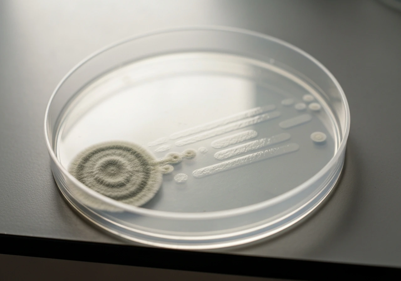
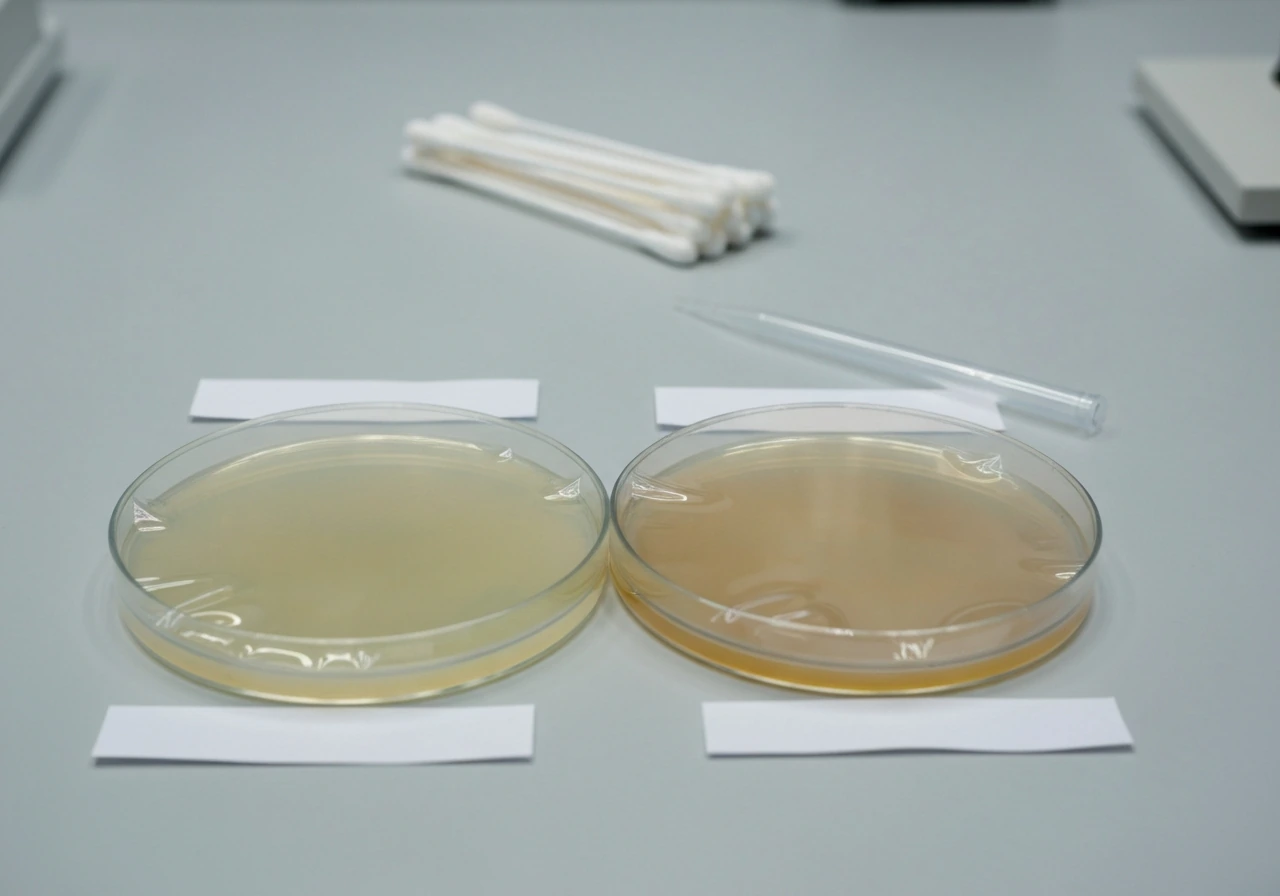
What fungal colonies look like compared to bacterial growth
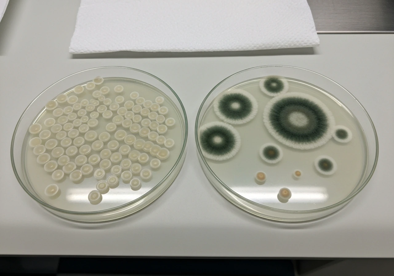
This is usually where people first realize something is off. Bacterial colonies on nutrient agar tend to be smooth, discrete, and relatively uniform. Fungal colonies look distinctly different, though it depends on whether you are dealing with a mold or a yeast.
| Feature | Mold colonies | Yeast colonies | Typical bacterial colonies |
|---|---|---|---|
| Texture | Fluffy, cottony, woolly, or powdery | Smooth, creamy, or pasty | Smooth, glistening, or mucoid |
| Appearance | Filamentous, spreading edges | Compact, rounded | Compact, defined edges |
| Color | White, green, black, gray, or other pigments | White, cream, or off-white | Usually white, cream, or yellow |
| Speed of growth | Slower (may appear day 2–6) | Moderate (24–48 hours or more) | Fast (often 18–24 hours) |
| Surface spread | Spreads outward with fuzzy margin | Stays compact | Stays compact or spreads smoothly |
The most reliable visual flag for a mold is the fuzzy, aerial mycelium radiating outward from a central point. If you see something that looks like a tiny cotton ball or a dusting of powder spreading across the agar, that is almost certainly a mold colony. Yeasts can be trickier because their colonies look superficially similar to bacterial colonies. They tend to be slightly larger, sometimes wetter or more opaque, and may have a subtle doughy or yeasty smell. The key takeaway: if any colony looks fuzzy, filamentous, or shows color pigmentation like green or black, treat it as a likely fungal contaminant immediately.
How to quickly confirm whether you are looking at a fungus
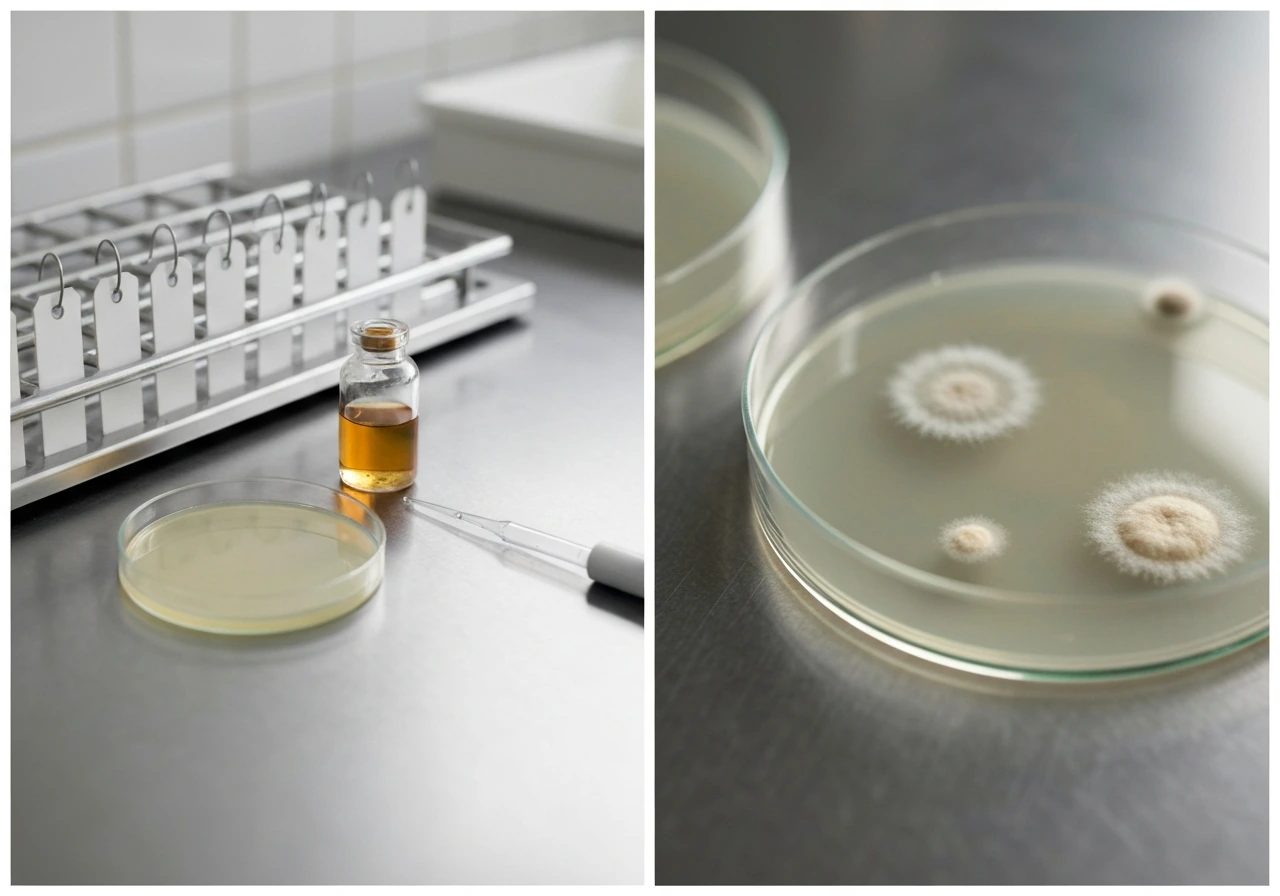
Visual morphology gets you most of the way there, but if you need confirmation or if the colony looks ambiguous, a simple wet mount under a microscope is the fastest approach. Scrape a small amount of material from the suspect colony using a loop or needle, place it in a drop of water on a slide, and look for hyphae, pseudohyphae, or spores. Hyphae are branching filamentous structures that bacteria simply do not have. Hyphae are able to grow and penetrate tissue, which is why fungal identification matters for preventing spread the hyphae are able to grow and penetrate. If you see them, you have a fungus.
For a more definitive stain, lactophenol cotton blue (LPCB) is the standard. It binds to chitin in fungal cell walls and stains hyphal structures blue, making identification under the microscope much cleaner. You can also use a KOH wet mount preparation, which helps clear background material and makes fungal elements easier to see, especially for yeasts showing budding or pseudohyphae. Neither technique requires specialized equipment beyond a basic light microscope.
If you do not have microscopy available, the practical rule is this: if a colony appeared later than your bacteria (after 48 to 72 hours), looks fuzzy or pigmented, or is spreading in a way inconsistent with your expected organisms, assume it is a fungal contaminant and act accordingly.
What to do with a contaminated plate and how to avoid it next time
If your plate has visible fungal growth, the most important thing is to stop using it for your experiment. Mold colonies on agar can spread aggressively and release spores, which will contaminate adjacent plates, your bench, and potentially your incubator. Seal the plate with tape before moving it, and discard it through your lab's standard biohazard waste process. Do not open the plate unnecessarily once significant mold growth is visible.
For prevention going forward, start with your aseptic technique. Keep the lid over the plate as much as possible when working, minimize the time plates are open to air, and avoid working near air vents or areas with a lot of foot traffic. Airborne spores are everywhere, and the less time your agar is exposed, the better.
- Store poured plates sealed (parafilm or plastic bags) and refrigerated at 4°C until use.
- Invert plates during incubation to reduce condensation dripping onto the agar surface.
- Use plates within a reasonable shelf life (typically 4 to 6 weeks for commercially prepared plates, less for homemade).
- Wipe down your bench with 70% ethanol before working and allow it to dry.
- Work close to a flame or inside a biosafety cabinet if available.
- Incubate at 35 to 37°C if your target bacteria can tolerate it, since this temperature range is less favorable for many environmental molds.
- Check plates at 24 and 48 hours. Remove and discard any plate showing early fungal growth before spores can spread.
Environmental context matters too. In food safety work, the presence of airborne fungal spores is directly tied to the cleanliness of the surrounding environment. High-humidity rooms, water-damaged materials nearby, and heavy organic dust all increase spore loads in the air. This is the same principle underlying why mold growth in food processing environments is so tightly controlled. The agar plate is just a detector for whatever is in your lab environment.
How to inhibit fungal growth when you are trying to culture bacteria
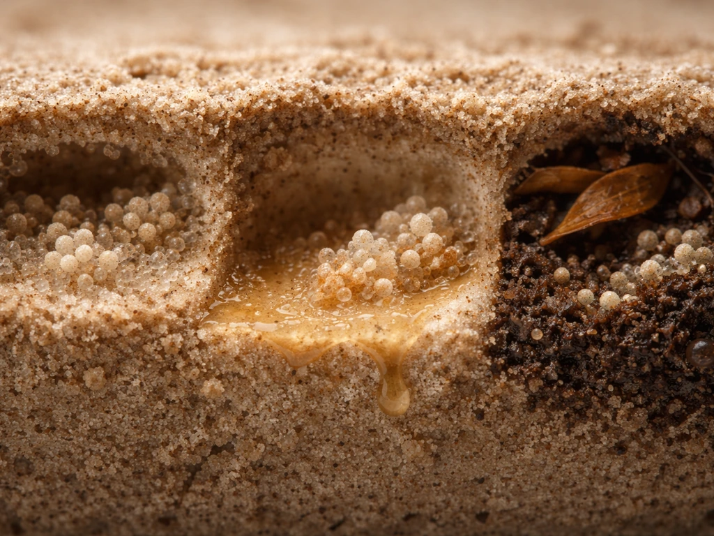
If fungal contamination is a recurring problem, the most effective solution is switching to a more selective medium rather than fighting it with technique alone. Nutrient agar is simply not designed to resist fungal colonization. In contrast, fungus generally does not grow inside an ant colony because the ants’ hygiene behavior and antimicrobial defenses help keep microbial growth in check why does the fungus not grow inside the ant colony. Here are your main options:
- Switch to a selective or differential medium appropriate for your target bacteria. Many bacterial selective media contain antibiotics or bile salts that do not affect your bacteria but will slow or stop fungal growth.
- Add an antifungal to your existing medium formulation. Cycloheximide (actidione) is commonly used in mycology media to suppress certain molds, and chloramphenicol is used in SDA formulations to suppress bacteria, so the reverse logic applies: antifungals can be added to bacterial media to suppress fungi. Check compatibility with your target organisms first.
- Acidify your medium. Most human-pathogenic and food-relevant bacteria tolerate a pH around 6.5 to 7.0, but lowering pH toward 5.5 to 6.0 (as done in Sabouraud formulations to suppress bacteria) also works in reverse: bacteria-favoring media can be made slightly less hospitable to acid-sensitive molds, though this approach has limits and may affect your bacteria too.
- Tighten your incubation conditions. Incubate at 35 to 37°C and read plates early (18 to 24 hours). This window lets fast-growing bacteria develop while giving slow-growing environmental molds less time to establish.
- For environmental monitoring or food safety testing where fungal contamination is expected to be high, consider using two parallel plates: one standard nutrient agar for bacteria and one fungal-specific medium (like potato dextrose agar or SDA) to enumerate fungi separately. This separation is standard practice in air and surface monitoring programs.
Potato dextrose agar (PDA) is worth mentioning here as a point of comparison: it can be acidified or supplemented with chloramphenicol specifically to suppress bacteria while cultivating fungi. That is the fungal equivalent of what you are trying to achieve in reverse. The principle is the same: use pH, selective antibiotics, or both to create a medium that favors one group over the other. Nutrient agar does neither, which is why it remains vulnerable to fungal overgrowth when conditions allow.
The bottom line is that fungal contamination on nutrient agar is common, recognizable, and manageable. You can usually identify it within 48 to 72 hours by colony morphology, confirm it quickly with a basic wet mount, and prevent it with better aseptic technique or a more selective medium. If you are seeing it repeatedly, that is your cue to investigate your environment for moisture and spore sources, not just your bench technique. Resident flora typically grow where they can access moisture and nutrients, such as on the skin surface or in the body’s mucosal environments where does resident flora grow.
FAQ
If my plate was incubated only 24 hours, can fungal growth still appear later?
Yes. Many molds are slower than common bacteria, so a “clean” 24-hour check can turn into noticeable fungal colonies after 3 to 6 days, especially at 20 to 25°C. If you are expecting bacteria, include a longer secondary read before you discard control plates.
Does nutrient agar support yeast and molds equally?
Both can grow, but they behave differently. Yeast often starts showing colonies sooner and may look smooth, creamy, or slightly opaque. Molds usually form fuzzy, filamentous mycelium and pigment. If you see spreading fuzz, treat it as mold even if bacteria were the target.
Can condensation alone cause fungi to grow even if my technique was good?
Condensation can provide the extra free moisture spores need to germinate quickly. Temperature swings, storing plates upright when they were previously warm, and high humidity in the incubator all increase wet surface conditions. In practice, control storage (sealed, refrigerated when appropriate) and incubator humidity to reduce this trigger.
What should I do if I suspect fungal contamination but the colonies look borderline?
Do not rely only on “mostly smooth” appearance. If timing is inconsistent with your expected organisms, or if the colony edge seems irregular or shows any pigment, confirm with a wet mount or LPCB staining. Also, keep the plate sealed and isolated until you decide, since ambiguous colonies can still be sporulating.
Is it safe to smell plates to decide whether it is yeast?
Avoid sniffing open plates. Odors can be misleading, and some contaminants are hazardous. If your lab uses an odor-based observation, it should be done only from sealed plates and with appropriate biosafety procedures, otherwise use microscope confirmation.
How can I tell whether a colony is growing because it is truly on the agar or just due to surface wetness?
Look for growth that extends from a point of contact and shows stable colony morphology over time. Surface droplets can spread material, making colonies look smeared. With moisture-related smearing, the pattern usually lacks the organized radial fuzz, pigmentation, or consistent colony growth you would expect from a real fungal colony.
If fungal colonies show up, should I incubate the plate longer to see if it stops?
Usually no. Leaving plates longer increases sporulation risk and cross-contamination. Instead, stop using the plate for experiments, seal it for disposal, and confirm identity only if needed via a small, controlled diagnostic workflow following your lab safety rules.
Will incubating at 35 to 37°C reliably prevent fungi on nutrient agar?
It reduces the growth of many environmental fungi, but it does not guarantee prevention. Some molds, including certain Aspergillus species, can tolerate warmer temperatures. If fungal contamination is recurring, rely on selection (like pH control or antibiotics in a fungus-favoring medium) rather than temperature alone.
If I switch to a selective medium, does that eliminate the need for aseptic technique?
No. Selective media only tilt the odds. You can still get breakthrough fungi from heavy spore loads, and selection can sometimes suppress bacteria while allowing unexpected organisms to grow. Continue minimizing plate exposure time, reducing drafts and dust, and sealing plates immediately after work.
Could fungal contaminants come from my water, reagents, or glove contact rather than airborne spores?
Yes. Contaminated diluent water, improvised wet mounts, dusty gloves, and residue on work surfaces can introduce spores or living cells. If you keep seeing repeat contamination patterns at the same spots or on all plates from the same batch, consider reagent and consumable sources in addition to air and condensation.
Are colonies on the negative control plates an expected outcome?
If your negative controls are truly uninoculated, visible colonies usually indicate contamination from technique, environment, or incubation conditions. A “few late” colonies can still be informative, but if you routinely see fungal growth on blanks, it is a sign to audit workflow, humidity, and handling practices.
Next Article
Locations Where Yeast Might Naturally Grow and Why
Find real locations yeast can grow naturally, and learn the moisture, pH, food, and temperature conditions that enable i