The bacteria you most reliably expect to grow and produce a positive blackening reaction on bile esculin agar (BEA) are enterococci (Enterococcus faecalis, Enterococcus faecium, and related species) along with group D streptococci such as Streptococcus bovis. These organisms can tolerate bile and hydrolyze esculin, which turns the agar dark brown to jet black. A handful of other organisms, including Listeria monocytogenes, some Aerococcus species, and occasional other gram-positives, can also give positive or partial reactions, which is why BEA is described as presumptive, not definitive. E. coli has its own optimal growth temperature, so confirmatory testing and correct culturing conditions matter when you suspect it E. coli optimal growth temperature.
What Bacteria Grow on Bile Esculin Agar and How to Read It

What bile esculin agar is actually for
BEA is a selective and differential medium, meaning it does two jobs at once. It filters out a wide range of organisms that cannot survive in the presence of bile, and it reveals which survivors can hydrolyze esculin by producing a visible color change. The primary historical use has been presumptive identification of group D streptococci and enterococci from clinical specimens, food samples, and pharmaceutical products. In food microbiology, the FDA's Bacteriological Analytical Manual (BAM) references BEA formulations (Media M18) specifically for identifying these organisms in food matrices.
The test logic is straightforward: if something grows on BEA and the agar around it turns black, the organism passes both hurdles. It tolerates bile salts and it makes the enzyme that breaks esculin down. That two-part result is what gives you the presumptive identification.
How the medium selects and differentiates
There are three key components doing the work here. The bile (usually Oxgall, at around 40 g per liter in common formulations) acts as the selective pressure. Bile is a natural detergent that disrupts the cell membranes of many gram-positive organisms and most gram-negative organisms that are not adapted to it. Organisms that normally colonize the intestinal tract have evolved tolerance to bile, so they grow while most environmental contaminants and non-enteric bacteria do not.
Esculin (around 1 g/L) is the substrate. FDA’s BAM Media M18 (Bile Esculin Agar) specifies components such as esculin (1 g) and ferric citrate (0.5 g), supporting presumptive esculin hydrolysis with ferric-ion blackening blank" rel="noopener noreferrer">Esculin (around 1 g/L) is the substrate. When an organism produces the enzyme esculinase (a beta-glucosidase), it cleaves esculin into glucose and esculetin. Esculetin is the important product here because it immediately reacts with ferric citrate (around 0.5 g/L) in the medium to form an insoluble, dark iron-phenolic complex. That complex is the black precipitate you see spreading through the agar. The final pH of the medium sits at about 6. Hardy Diagnostics’ bile esculin agar formulation lists Oxbile (Oxgall) at 40.0 g/L, Esculin at 1.0 g/L, and ferric citrate at 0.5 g/L, with blank" rel="noopener noreferrer">a final pH of 6.6 ± 0.2 at 25°C. Some bacteria, including E. coli, can grow under certain acidic conditions, but growth depends strongly on the exact pH and exposure time can e coli grow in acidic environment. 6, which supports the growth of these enteric-associated organisms while adding one more subtle layer of selectivity.
The bacteria that grow on bile esculin agar
The classic positives are enterococci and group D streptococci. Here is a practical breakdown of the main groups:
| Organism | BEA Growth | Blackening Reaction | Notes |
|---|---|---|---|
| Enterococcus faecalis | Strong | Strongly positive (jet black) | Most reliable positive; common in food and clinical samples |
| Enterococcus faecium | Strong | Strongly positive | Second most common Enterococcus; clinically significant |
| Other Enterococcus spp. (E. avium, E. durans, E. gallinarum) | Variable to strong | Positive | Most species are bile-esculin positive |
| Streptococcus bovis group (S. gallolyticus, S. equinus) | Good growth | Positive | Classic group D strep; important in clinical ID |
| Listeria monocytogenes | Grows | Positive (black) | Key food safety concern; positive on BEA but distinct colony morphology |
| Aerococcus viridans | Variable | Weak to positive | Occasional positive; usually ruled out by other tests |
| Some Leuconostoc spp. | Limited | Weak or negative | Generally inhibited; rare weak reactions reported |
The standout from a food safety perspective is Listeria monocytogenes. It is bile-tolerant and esculin-positive, so it will grow and blacken BEA. This is actually used intentionally in some Listeria-selective media that are built on a BEA base. In food microbiology workflows, encountering a black-positive colony on BEA from a food sample should prompt you to think about both enterococci and Listeria until you run confirmatory tests. Cetrimide agar is also selective for certain gram-negative bacteria such as Pseudomonas aeruginosa, which may grow on it depending on the formulation and incubation conditions.
What to look for: reading growth and blackening
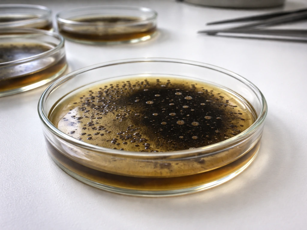
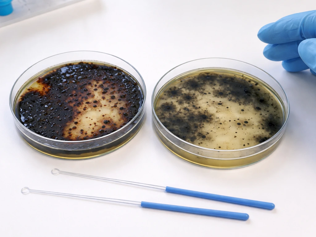
After incubating plates at 35 to 37°C for 18 to 24 hours, you are looking for two things simultaneously: actual colony growth and the black halo or darkening spreading into the agar around and beneath the colonies.
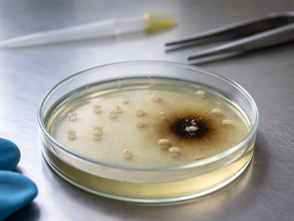
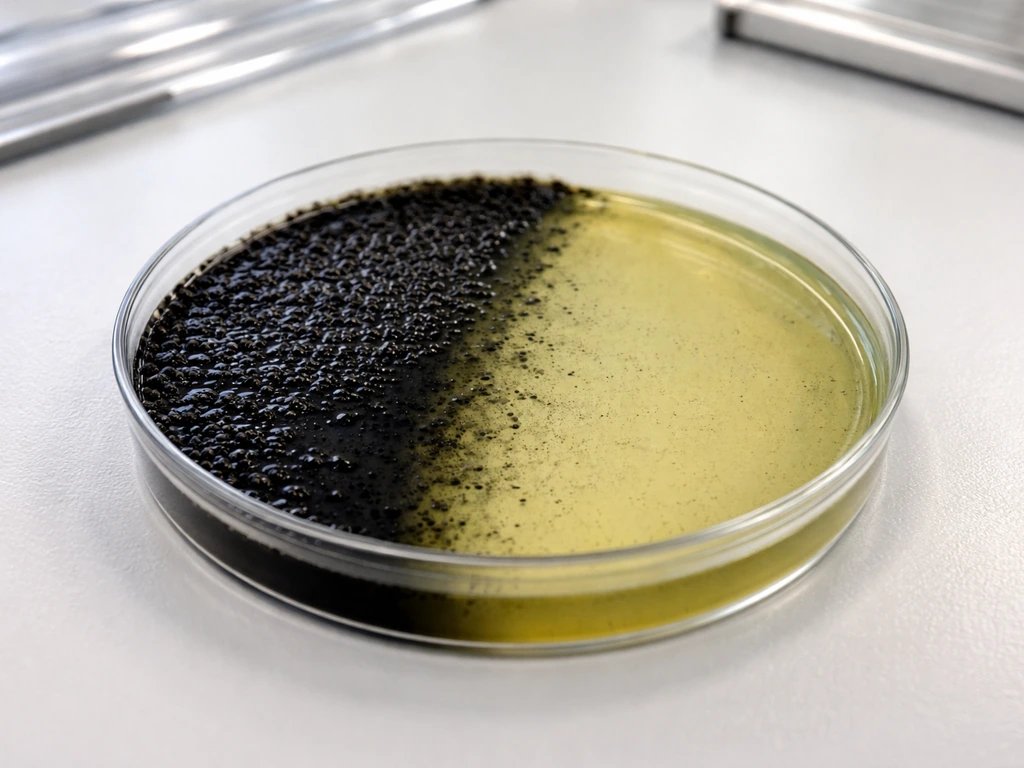
- A positive result is growth plus at least half the agar surrounding the colony turning dark brown to jet black. Many labs require greater than 50% blackening of the zone around a colony to call it positive.
- A negative result is either no growth or growth without any blackening. Some organisms can survive the bile (partial tolerance) but cannot hydrolyze esculin, giving you colonies with no color change.
- A weak or equivocal result is slight browning without definitive black. Do not call this positive. Extend incubation to 40 to 48 hours and reassess.
- The blackening is agar-diffusible. It spreads beyond the colony itself, which is one visual cue that distinguishes a true enzymatic reaction from surface pigment produced by the organism.
Enterococcus colonies on BEA are typically small to medium, gray-white to slightly brown, with a surrounding black zone that can become so pronounced the entire agar surface in that area turns black with heavy inocula. Listeria colonies tend to be smaller and smoother, and the blackening is similarly robust.
Look-alikes and how to tell them apart
The biggest identification challenge is separating enterococci from other bile-esculin–positive organisms, particularly when a BEA-positive result from a food or clinical sample could represent any of several genera. Here are the most common look-alikes and the tests that separate them:
| Organism | BEA Result | Key Differentiating Tests | Expected Outcome |
|---|---|---|---|
| Enterococcus spp. | Positive (black) | PYR test, 6.5% NaCl broth, growth at 10°C and 45°C | PYR positive, grows in 6.5% NaCl, grows at both temps |
| Streptococcus bovis group | Positive (black) | PYR test, 6.5% NaCl broth, Lancefield grouping | PYR negative, does NOT grow in 6.5% NaCl, group D antigen |
| Listeria monocytogenes | Positive (black) | Catalase test, Gram stain, motility at 25°C, hemolysis on blood agar | Catalase positive, small gram-positive rod, tumbling motility, beta-hemolytic (CAMP test) |
| Aerococcus viridans | Weak positive | Catalase, tetrad arrangement on Gram stain, PYR | Catalase negative, tetrads, PYR positive but growth in 6.5% NaCl is variable |
| Pediococcus spp. | Occasional positive | Gram stain morphology (tetrads), gas from glucose, vancomycin susceptibility | Tetrads, no gas from glucose, often vancomycin resistant |
In practice, the PYR test (pyrrolidonyl arylamidase) and growth in 6.5% NaCl broth together are the fastest ways to confirm Enterococcus. A BEA-positive colony that is also PYR-positive and grows in 6.5% NaCl is Enterococcus until proven otherwise. Adding a catalase test immediately separates Listeria (catalase positive, rod-shaped) from enterococci (catalase negative, cocci).
This matters especially in food microbiology because enterococci and Listeria carry very different contamination implications for a food facility or clinical investigator. Confusing them based on BEA alone is a real risk if you stop at the presumptive step. This parallels the situation with other selective media, where similar selectivity principles mean you always need a second confirmatory layer before reporting a result with confidence. Ideonella sakaiensis is different from the classic BEA positives, so a reported match should be confirmed with additional identification tests beyond growth and blackening on the medium other selective media.
A practical workflow you can follow today
Preparation and inoculation
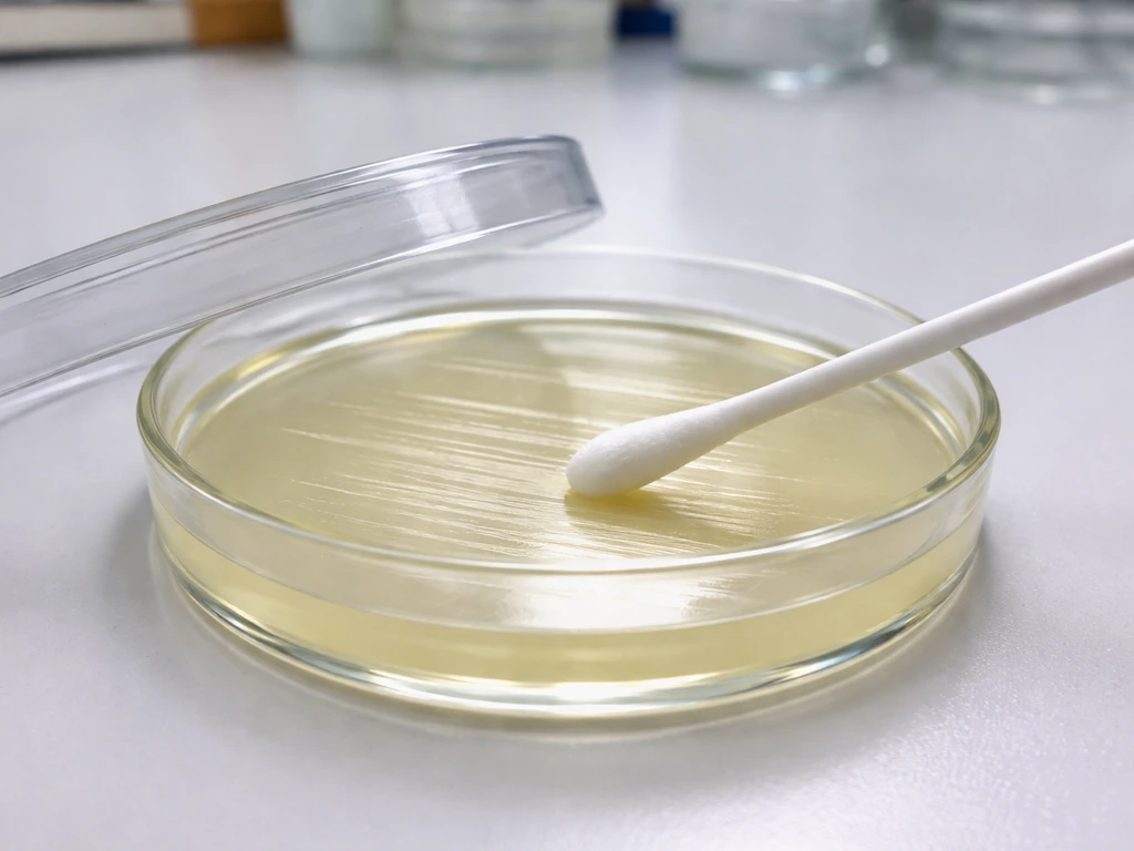
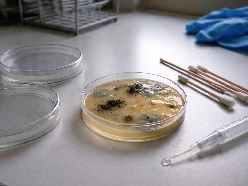
- Use freshly prepared or properly stored BEA plates. Plates stored longer than two to three weeks at 4°C can have degraded esculin or dried surfaces, both of which affect results. Check that the agar is slightly yellow-amber before inoculation.
- For pure culture testing, streak a single well-isolated colony directly onto the BEA surface using a standard loop. For food or environmental samples, inoculate from enrichment broth (for example, a bile-containing enrichment for Listeria or enterococcal enrichment broth) onto BEA as a secondary plating step.
- If you are testing multiple isolates, streak individual quadrants or use separate plates. Cross-contamination between strongly positive and weakly reactive isolates can cause false impressions of positivity.
Incubation
- Incubate at 35 to 37°C for 18 to 24 hours. This is the standard window; most enterococci produce visible blackening within this period.
- For samples where growth is slow or the inoculum is small, extend incubation to 40 to 48 hours before reading a final result. Do not call a plate negative at 18 hours if growth is present but blackening is still developing.
- Incubate in ambient air (aerobic conditions). BEA does not require CO2 enrichment for the target organisms.
Reading results
- Examine plates in good light. Look for the black precipitate in the agar, not just on the colony surface.
- Record both growth and blackening. An organism that grows without blackening is not bile-esculin positive.
- Pick suspicious colonies for confirmatory testing immediately. Subculture to blood agar or non-selective agar for Gram stain and secondary tests.
Pitfalls and troubleshooting
BEA is reliable, but a few consistent problems show up in real laboratory work. Knowing them in advance saves time.
- False positives from Listeria in food samples: If you are not expecting Listeria and do not run catalase or check morphology, you will misidentify it as Enterococcus. Always Gram stain your BEA-positive colonies from food samples.
- Weak positives from non-target organisms: Occasional strains of Leuconostoc, Pediococcus, or even some gram-negative organisms with unusual bile tolerance can produce faint darkening. These are rarely mistaken for true positives if you require robust blackening of at least 50% of the surrounding zone.
- Over-incubation artifacts: Plates incubated beyond 72 hours can show non-specific darkening from media oxidation or nutrient exhaustion, not esculin hydrolysis. Read within 48 hours for reliable results.
- Dried or cracked agar: Plates that have dried out do not allow the diffusion of esculetin-iron complexes properly. The black reaction may be confined directly under the colony and look less convincing. Retest on fresh plates.
- False negatives from inhibited inoculum: If your starting culture is stressed (from harsh enrichment conditions, freezing, or heavy antibiotic exposure), growth on BEA may be slow or absent even for true enterococci. Use a subculture on non-selective agar first to revitalize before BEA plating.
- Inoculum size effects: Too-heavy inoculum can cause entire plate blackening quickly, masking the differential interpretation. Use well-isolated colonies or diluted inocula.
What BEA-positive results mean in the real world
In food safety, finding bile-esculin–positive organisms in a food product or food-contact environment carries specific implications depending on which organism is confirmed. If you are instead working with mannitol salt agar, you would use its specific indicators to determine whether E. If you want to know whether E. coli will grow on a TSA plate, you should consider its general ability to grow on nonselective media like TSA. coli can grow there bile-esculin–positive organisms. Enterococci are considered indicator organisms in some contexts, meaning their presence suggests possible fecal contamination or sanitation failures. They are not always direct pathogens, but their presence in a ready-to-eat food at high counts is a regulatory concern in many frameworks.
Listeria monocytogenes on BEA from a food or environmental swab is a much more serious finding. Listeria is a foodborne pathogen with significant public health implications, and a presumptive positive from BEA must be confirmed and acted on promptly. This is one reason why food microbiologists treating BEA as a quick presumptive screen should always follow through with confirmatory identification.
In clinical microbiology, enterococcal identification from patient samples (urine, wound, bloodstream) has direct treatment implications given the increasing prevalence of vancomycin-resistant enterococci (VRE). A bile-esculin positive, PYR-positive, salt-tolerant gram-positive coccus from a clinical source warrants full species-level identification and susceptibility testing.
From a safe handling standpoint: treat all cultures on BEA as potentially pathogenic until confirmed otherwise. Enterococci and especially Listeria can persist on surfaces and equipment. Decontaminate plates and materials per your biosafety protocols before disposal, and avoid working with open plates outside a biosafety cabinet when processing food or clinical samples of unknown identity.
The bottom line is that BEA is a powerful first-step tool, but the black agar is the beginning of your identification, not the end. Pair it with PYR, salt tolerance, catalase, and a Gram stain and you will have a clear picture of what you are actually dealing with.
FAQ
If a plate shows no blackening on bile esculin agar, does that mean no target bacteria are present?
Not necessarily. Some organisms may grow poorly due to inoculum size, incubation time, or transport stress, so you can see weak growth without the full black halo. Also, partial reactions can occur, so re-check after the full recommended window and confirm with a suitable confirmatory step (for example, Gram stain plus the appropriate enzyme tests).
Can E. coli grow on bile esculin agar and still confuse the result?
Yes, E. coli can sometimes grow, particularly if incubation conditions allow it, but BEA requires both bile tolerance and esculin hydrolysis for a convincing blackening pattern. If you suspect enteric gram-negatives, rely on colony morphology and confirmatory identification rather than using blackening alone, since gram-negative growth without strong, spreading blackening can be misleading.
Why do some labs report a “partial” black reaction on BEA, and how should I interpret it?
Partial blackening often reflects weaker or slower esculin hydrolysis, lower enzyme activity, or limited ferric citrate interaction around the colony. Treat partial positives as presumptive, repeat reading at the end of the incubation period, and then confirm with PYR for presumptive Enterococcus and a catalase check to screen out Listeria.
What should I do if the colonies are present but the blackening stays very localized to the colony edge?
That pattern suggests limited diffusion of the iron-phenolic complex, which can happen with low inoculum or borderline esculinase producers. Don’t jump to identification from the halo size alone. Use a Gram stain and then the rapid discriminators (PYR and salt tolerance for Enterococcus, catalase for Listeria) to decide the next steps.
How do I avoid mixing up Enterococcus and Listeria when both can blacken BEA?
Use colony characteristics plus at least one rapid differential test. A common practical sequence is: Gram stain first, then PYR and 6.5% NaCl broth for Enterococcus, and catalase for Listeria. Enterococcus is typically PYR-positive and salt tolerant, Listeria is catalase positive and classically shows rod morphology, which reduces false presumptive calls.
Does BEA require the exact incubation temperature and time to be reliable?
Yes. Deviations can change whether marginal esculin hydrolysis is detected and can also alter the balance between bile-tolerant organisms and background survivors. Stick to the standard incubation range and complete the full reading window, and if you must change conditions (for example, shorter holds), you should treat results as more presumptive and confirm anyway.
How should I handle BEA plates from food versus clinical specimens differently?
The medium reading logic is the same, but the confirmation threshold is higher for clinical work and regulatory decisions in food safety. For food/environmental samples, you should still confirm presumptive BEA positives promptly because Listeria carry different actions than enterococci. For clinical specimens, proceed to full species-level identification and, when indicated, antimicrobial susceptibility testing due to resistance concerns.
Can I rely on colony color (gray-white versus brown) instead of confirmatory tests?
Not by itself. Color shades can vary with inoculum density, medium batch differences, and incubation microconditions. BEA’s key signal is the blackening from esculin hydrolysis, but final identification should still be anchored to confirmatory tests like PYR, catalase, and salt tolerance, plus Gram stain.
What biosafety and disposal steps should I follow after a presumptive BEA positive?
Treat presumptive positives as potentially pathogenic until ruled out, especially for Listeria and resistant enterococci. Keep plates contained, minimize aerosol-generating actions, and dispose of materials according to your facility’s biosafety procedures for unknown enteric-like isolates.
If my organism is BEA-positive but doesn’t fit the expected Enterococcus or Listeria pattern, what’s the best next step?
Don’t stop at BEA. Move to broader identification, such as systematic biochemical profiling or an accepted identification platform, and include a targeted differential panel (for example, catalase, PYR, and salt tolerance) alongside Gram stain. This is especially important when the isolate shows atypical morphology or inconsistent blackening.
Will E. coli Grow on a TSA Plate? Growth Conditions and Timing
Yes, E. coli typically grows on TSA at 35–37°C within 18–24h; includes conditions, timing, and confirmation tips.