Cetrimide agar is designed specifically to grow Pseudomonas aeruginosa while suppressing nearly everything else. That is the short version. If you see growth on cetrimide agar, your first assumption should be P. aeruginosa, but there are a handful of other Pseudomonas species and occasional non-Pseudomonas organisms that can push through depending on incubation conditions, inoculum load, and the source material. This guide walks through what to expect, what looks suspicious, and what to do next if your result is ambiguous. Pseudomonas aeruginosa and other gram-negative bacteria can behave very differently across pH ranges, so if you are also thinking about survival rather than just selective growth, you may want to review whether can e coli grow in acidic environment.
What Bacteria Can Grow on Cetrimide Agar and How to Confirm

What cetrimide agar is actually doing
Cetrimide agar is a solid selective medium. The active ingredient is cetrimide (cetyltrimethylammonium bromide), a quaternary ammonium compound that disrupts cell membranes and kills or inhibits the vast majority of gram-positive and gram-negative bacteria. P. aeruginosa tolerates cetrimide because it has robust outer membrane resistance mechanisms that most other organisms lack.
A typical formulation (based on Sigma-Aldrich's cetrimide agar base) contains around 0.5 g/L cetrimide, 20.5 g/L peptone as a nutrient source, 1.4 g/L magnesium chloride, 10.0 g/L potassium sulphate, and 13.6 g/L agar, with a final pH of 7.2 plus or minus 0.2. The magnesium chloride and potassium sulphate are not just fillers: they actively stimulate P. aeruginosa to produce its characteristic pigments, pyocyanin (blue-green) and pyoverdine (yellow-green fluorescent). This is what makes cetrimide agar both selective and differential at the same time. The FDA's Bacteriological Analytical Manual (BAM) lists it as medium M37 for exactly this purpose: isolating and identifying P. What bacteria grow on bile esculin agar depends on the medium's selectivity for bile-tolerant, esculin-hydrolyzing organisms medium M37. aeruginosa.
Bacteria that are expected to grow
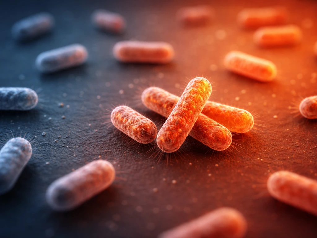
Pseudomonas aeruginosa is the primary target and the most common organism you will find on cetrimide agar. It grows well, typically producing colonies in the 0.5 to 3.0 mm range with a straw to greenish coloration and often visible blue-green pigmentation spreading into the agar. Thermo Fisher's instructions for use describe the positive control appearance (P. aeruginosa ATCC 10145) as straw-colored colonies with green pigmentation. You may also notice a fruity, grape-like odor, which is one of P. aeruginosa's well-known sensory characteristics.
Beyond P. aeruginosa, several other Pseudomonas species can show growth on cetrimide agar, though usually with less vigorous results. Pseudomonas fluorescens and members of the P. fluorescens complex may grow and produce yellow-green fluorescence but will not produce pyocyanin. Pseudomonas putida is another occasional grower. These species share enough of the outer membrane resistance profile to tolerate cetrimide at the concentrations used in standard formulations. They are considered partial or breakthrough growth rather than target growth, and distinguishing them from P. aeruginosa requires follow-up testing.
Other organisms that may show up (and why this matters)
Cetrimide agar is highly selective but not perfectly exclusive. In practice, a few non-Pseudomonas organisms can occasionally break through, particularly when the inoculum is very heavy or the source material is complex. Burkholderia cepacia complex organisms are the most clinically significant example: they share some cetrimide tolerance with Pseudomonas and can grow on cetrimide agar, which creates a real identification problem in clinical and environmental microbiology settings. Some Acinetobacter strains and certain Stenotrophomonas maltophilia isolates have also been documented as occasional cetrimide agar growers under specific conditions.
On the flip side, strongly cetrimide-susceptible organisms like Enterococcus faecalis are expected to show no growth at all. Cetrimide agar is not a standard medium for assessing whether E. coli will grow on a TSA plate, so those conditions should not be assumed to translate directly. Thermo Fisher's IFU explicitly lists E. faecalis ATCC 29212 as a negative control organism, confirming that gram-positive cocci should be fully inhibited. If you see anything resembling gram-positive-type colonies, that is a contamination signal or a medium integrity issue, not a true positive.
Mixed cultures from environmental samples (water, soil, clinical wounds, food processing surfaces) carry the most risk of confusing results. A mixed plate can show P. aeruginosa's characteristic blue-green colonies alongside pale, non-pigmented colonies from P. fluorescens complex species, which is exactly the interpretive complication described in Clinical Microbiology Reviews. Do not assume every colony on a cetrimide plate is P. If your goal is to evaluate how Ideonella sakaiensis grows, plate-based conditions and the incubation setup should be tailored to its specific growth requirements rather than assuming it will follow Pseudomonas-like behavior. aeruginosa.
How incubation conditions change what you see
Temperature and time matter more than most people realize with cetrimide agar. The standard incubation conditions are 36 plus or minus 2 degrees Celsius (so 34 to 38 degrees Celsius) under aerobic conditions for 40 to 48 hours, according to Thermo Fisher's IFU. Some protocols reference 35 to 37 degrees Celsius with evaluation starting at 24 hours, and Merck's product sheet uses 35 to 37 degrees Celsius as the target range. The difference between 30 degrees Celsius and 37 degrees Celsius incubation has measurable effects on pigment production: research comparing multiple time points (24, 48, and 72 hours) at different temperatures shows that pyocyanin and pyoverdine output varies significantly based on both variables.
P. aeruginosa grows optimally at 37 degrees Celsius and can tolerate up to 42 degrees Celsius, which is actually used as a selective feature in some identification schemes. Incubation temperatures for E. coli are typically different from the settings used for cetrimide agar targeting Pseudomonas 42 degrees Celsius. Organisms like P. fluorescens prefer cooler temperatures (around 25 to 30 degrees Celsius) and grow poorly or not at all at 42 degrees Celsius. Running a second plate at 42 degrees Celsius alongside your standard 37 degrees Celsius plate is a simple and practical way to get additional differentiation data with no extra reagents.
Oxygen availability is also critical. Cetrimide agar should be incubated aerobically. P. aeruginosa is an obligate aerobe for practical purposes (it can use nitrate as a terminal electron acceptor under low-oxygen conditions, but pigment production and robust growth require good oxygen availability). Placing plates in an anaerobic or microaerophilic environment will suppress growth and pigment even from a confirmed P. aeruginosa strain.
Interpreting results: what normal looks like vs. what should raise questions
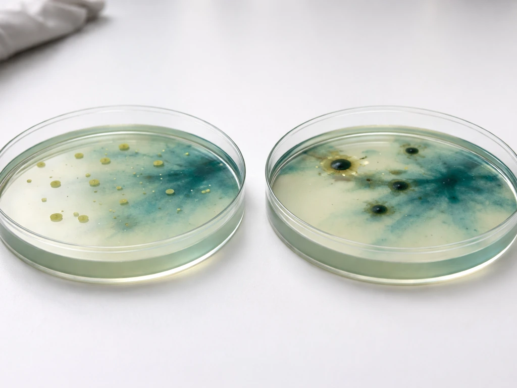
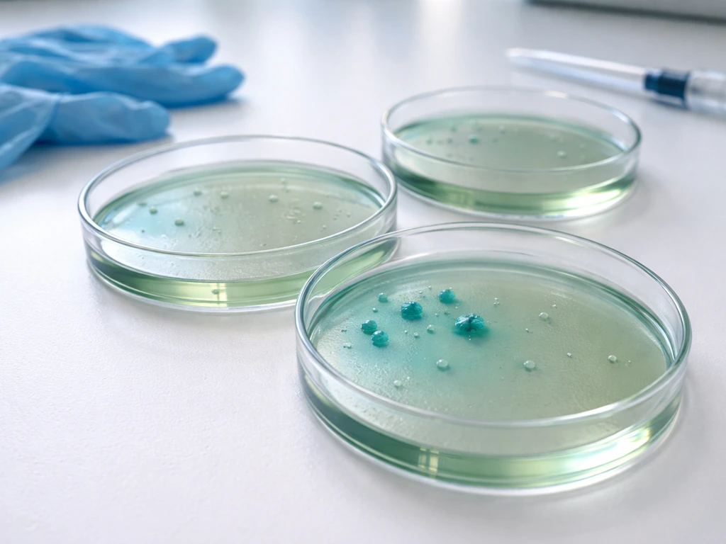
A clear positive result on cetrimide agar looks like this: colonies 0.5 to 3.0 mm in diameter, straw to greenish in color, with blue-green pigment diffusing into the agar, a fruity or grape-like odor, and growth that starts to be visible at 24 hours and is well-developed by 40 to 48 hours. That combination is strongly presumptive for P. aeruginosa.
Atypical results fall into a few categories worth knowing:
- White or pale colonies with no pigment: P. aeruginosa does not always produce pyocyanin or pyoverdine. Non-pigmented and mucoid strains are documented, particularly from cystic fibrosis patients. One JCM study found roughly 2% of P. aeruginosa strains failed to produce pigment under standard conditions. Do not rule out P. aeruginosa just because pigment is absent.
- Yellow-green fluorescence only, no blue pigment: Suggests P. fluorescens complex or another non-aeruginosa Pseudomonas. Pyoverdine without pyocyanin is a flag for further testing.
- Very small or weak colonies: May indicate a tolerant but not fully cetrimide-resistant organism, or an isolate under stress. Also seen with mucoid P. aeruginosa strains.
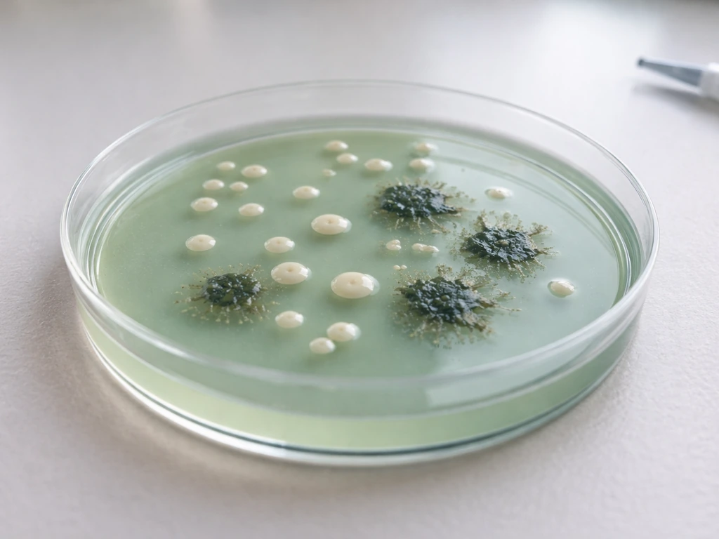
- Multiple colony morphologies on the same plate: A mixed inoculum from an environmental or clinical source. Treat each distinct morphology as a potential separate organism.
- No growth at all: Either the target organism was absent, the medium was incorrectly prepared, incubation conditions were wrong, or the inoculum was killed before plating.
Prolonged incubation beyond 48 to 72 hours can complicate interpretation because additional pigments accumulate and colony morphology spreads. If you need to extend incubation to 72 hours, document the appearance at 24 and 48 hours as well so you have a baseline.
Confirming your isolate: practical next steps
A positive or presumptive result on cetrimide agar alone is not enough to report P. aeruginosa. The standard first confirmatory step is an oxidase test. In a Reddit r/microbiology troubleshooting thread about identifying Pseudomonas aeruginosa on cetrimide agar, oxidase testing is also recommended as the next step when the plate appearance is ambiguous. P. aeruginosa is oxidase-positive, and this test takes about 30 seconds using a commercial oxidase strip or reagent. If your isolate is oxidase-negative, it is not P. aeruginosa, regardless of how it looks on cetrimide agar. This is the most commonly recommended immediate follow-up in practical microbiology settings, including the Reddit microbiology community's own troubleshooting discussions.
After confirming oxidase-positive status, the typical workflow for confirming P. aeruginosa identity includes the following steps:
- Gram stain: P. aeruginosa is a gram-negative rod. A gram-positive result immediately redirects identification.
- Growth at 42 degrees Celsius: Inoculate a blood agar or non-selective plate and incubate at 42 degrees Celsius for 24 hours. P. aeruginosa grows; P. fluorescens and most other Pseudomonas species do not.
- Pigment confirmation on King's A and King's B agar: King's A promotes pyocyanin production; King's B promotes pyoverdine fluorescence under UV light. These give you more controlled pigment data than cetrimide agar alone.
- Biochemical profile or API/VITEK: For definitive species-level ID, especially if atypical pigmentation or growth is observed, run a standard biochemical panel or automated ID system. P. aeruginosa has a characteristic profile including positive oxidase, glucose oxidation without fermentation, arginine dihydrolase positive, and negative for many sugars.
- Antibiogram or susceptibility testing: Relevant in clinical and food safety settings where you need to confirm the organism is P. aeruginosa and understand its resistance profile.
When working with environmental or food samples where the sibling question of whether organisms like E. For mannitol salt agar, the key question is whether a given organism can both tolerate the salt concentration and ferment mannitol. coli grow on selective media is also relevant (similar logic applies to mannitol salt agar for staphylococci or bile esculin agar for enterococci), the workflow principle is the same: selective agar gives you a presumptive population, and confirmatory tests give you identity. Never stop at the selective plate.
Troubleshooting when results don't make sense
No growth when you expected it
- Check medium preparation: Was the cetrimide concentration correct? Over-autoclaving can degrade cetrimide and also denature the agar structure. Follow the manufacturer's preparation instructions carefully.
- Confirm incubation temperature: Even a few degrees low (for example, 30 degrees Celsius instead of 36 to 37 degrees Celsius) can produce noticeably reduced or delayed growth.
- Check the inoculum: If you streaked from a refrigerated sample or a dry swab, the organism count may have been too low. Consider enrichment in a non-selective broth before plating.
- Verify aerobic conditions: Make sure the incubator is not accidentally running a reduced-oxygen environment.
Contamination or unexpected growth
- If you see dense, spreading growth of multiple colony types, your inoculum was too heavy or poorly mixed. Dilute and replate.
- If you see tiny pinpoint colonies that do not fit the P. aeruginosa description, they may be satellite growth from organisms that are partially inhibited but not fully suppressed. Pick and test separately before drawing any conclusions.
- Run your negative control (E. faecalis ATCC 29212 or equivalent) alongside every batch of new cetrimide agar. If your negative control shows growth, the medium is compromised and the batch should be discarded.
Weak or delayed pigment production
If colonies are growing but pigment is weak or absent, extend incubation to 48 to 72 hours and observe under both visible light and UV (365 nm) for pyoverdine fluorescence. Mucoid strains from chronic infection sources often produce little pigment. If pigment still does not appear, proceed directly to oxidase testing and biochemical confirmation rather than waiting on the plate. The absence of pigment does not rule out P. aeruginosa, it just means you cannot use pigment as your confirmatory feature.
A quick reference: expected growth patterns by organism
| Organism | Growth on Cetrimide Agar | Pigment | Notes |
|---|---|---|---|
| Pseudomonas aeruginosa | Good (expected positive) | Pyocyanin (blue-green) and/or pyoverdine (yellow-green fluorescent) | Primary target; some strains non-pigmented, especially mucoid CF isolates |
| P. fluorescens complex | Variable (partial grower) | Pyoverdine only (no pyocyanin) | Grows better at lower temperatures; no growth at 42°C helps differentiate |
| P. putida | Variable (weak to moderate) | May produce pyoverdine; no pyocyanin | Less common on cetrimide; confirm with oxidase and biochemicals |
| Burkholderia cepacia complex | Occasional breakthrough | None typical | Clinically significant; requires separate selective media for proper detection |
| Stenotrophomonas maltophilia | Rare breakthrough | None | Oxidase-negative; easy to rule out |
| Acinetobacter spp. | Rare breakthrough at high inoculum | None | Oxidase-negative; gram stain and biochemicals needed |
| Enterococcus faecalis | No growth (negative control) | None | Gram-positive; fully inhibited by cetrimide at standard concentrations |
Cetrimide agar is a well-established, reliable tool when used correctly. It narrows your suspect list to a small group of organisms, with P. aeruginosa as the clear primary target. The key is treating the plate result as the start of identification, not the end. Run your controls, confirm with oxidase and a temperature-tolerance test, and use biochemical profiling whenever the colony morphology or pigment pattern is anything less than textbook. If you are chasing an organism match beyond Pseudomonas results, it helps to also consider where does e coli grow as a related starting point for expected growth conditions and habitat.
FAQ
What bacteria can grow on cetrimide agar besides Pseudomonas aeruginosa?
Most growth will still be P. aeruginosa, but other Pseudomonas species (including members of the P. fluorescens complex) can show partial or breakthrough growth. Non-Pseudomonas organisms reported in some settings include members of the Burkholderia cepacia complex, certain Acinetobacter strains, and occasional Stenotrophomonas maltophilia isolates, especially with heavy inoculum or mixed samples.
If I see blue-green pigment, is that enough to confirm P. aeruginosa?
Not by itself. Pigment can be weak, absent in mucoid strains, or confusing in mixed plates. You still need confirmation, with oxidase testing being the fastest immediate check. Also verify pigment under both visible light and UV (about 365 nm) for pyoverdine.
Can non-Pseudomonas bacteria grow on cetrimide agar if I incubate longer than 48 hours?
Yes, prolonged incubation (beyond about 48 to 72 hours) can make interpretation harder and increase the chance of seeing additional breakthrough growth or pigment accumulation. If you extend incubation, record colony appearance at earlier time points (around 24 and 48 hours) so you can compare growth onset and morphology.
Why is growth on my cetrimide plate faint or different from expected?
Common causes include suboptimal oxygen conditions (plates should be incubated aerobically), incubation temperature outside the usual target range (roughly 34 to 38°C for standard workflows), and inoculum problems (very heavy or very mixed samples). Weak pigment can also happen with mucoid strains from chronic sources.
What should I do if colonies look gram-positive or “odd” for cetrimide agar results?
That is a contamination signal or a medium integrity/pre-analytical issue, not a true P. aeruginosa profile. Use Gram stain and verify your plate handling and controls, since cetrimide agar is meant to inhibit gram-positive organisms.
Can cetrimide agar be used to decide whether other organisms, like E. coli, can grow?
No. Lack of growth on cetrimide agar does not reliably predict performance on other media, because the selective pressure is specific to cetrimide tolerance and Pseudomonas membrane characteristics. If your goal is E. coli, you need appropriate media and conditions for that organism rather than extrapolating from cetrimide agar.
How can I differentiate P. aeruginosa from P. fluorescens complex colonies on the same type of plate?
A practical clue is that P. aeruginosa typically produces pyocyanin (often described as blue-green) while P. fluorescens complex organisms may show fluorescence without pyocyanin. Even so, confirm with oxidase and follow-up biochemical profiling when colony appearance or pigment patterns are not textbook.
What is a simple way to add differentiation using incubation temperature?
Run a second plate at a higher temperature (about 42°C) alongside your standard plate (around 35 to 37°C). P. aeruginosa can tolerate higher temperatures, while many P. fluorescens strains grow better at cooler temperatures (roughly 25 to 30°C).
Do I need a temperature-tolerance test after oxidase is positive?
Yes, as part of a confirmatory workflow. Oxidase positivity is necessary for typical presumptive identification, but temperature tolerance (and additional biochemical profiling when needed) helps resolve cases where organisms partially tolerate cetrimide or produce weak or atypical pigment.
What controls should I run with cetrimide agar to avoid misinterpretation?
Include at least one positive control strain (commonly P. aeruginosa ATCC-type strains used by vendors) and a negative control such as a known oxidase-negative gram-positive organism listed by the manufacturer. Controls help you distinguish true selectivity failures from issues like incorrect incubation conditions or compromised media.
What if my isolate is oxidase-negative but still grew on cetrimide agar?
Treat it as not P. aeruginosa regardless of appearance. Oxidase-negative isolates that grow are consistent with breakthrough organisms or mixed culture. Proceed with additional identification steps appropriate to the colony morphology and your sample source.
Can E. coli Grow in an Acidic Environment? pH Guide
Can E. coli grow in acidic foods? Learn pH limits, strain differences, and how acidity affects growth vs survival.