Several types of microorganisms can genuinely grow or replicate inside human lungs: bacteria, fungi, and certain parasites. Viruses are a different story since they need to hijack living host cells to reproduce and cannot grow independently in lung tissue the way a bacterium divides in a warm, moist environment. The distinction matters because the environmental conditions that allow a pathogen to establish itself in the lung, and the way a lab confirms its presence, depend entirely on what kind of organism you are dealing with.
Bugs That Grow in the Lungs: Pathogens, Niches, Controls

Marcus Reeves
11 Aug 2026
What we mean by 'growth' and 'replication' in the lung
These two words get used interchangeably, but they describe different biological processes. Growth, in microbiology, means an organism uses available nutrients, moisture, oxygen, and the right temperature to divide and increase its population independently. Bacteria and fungi do this. A bacterium like Streptococcus pneumoniae can take up nutrients from airway secretions and split into daughter cells without any help from the host's cellular machinery.
Replication refers specifically to how viruses reproduce. A virus injects its genetic material into a host cell and forces that cell to manufacture new viral particles. Outside a host cell, a virus cannot replicate at all. This is why viruses are called obligate intracellular parasites. The lung provides host cells for viruses to exploit, but the virus is not 'growing' in the environmental sense that a bacterium or mold would grow on a laboratory plate or in a food matrix.
Colonization and infection are also worth separating. Colonization means a microorganism establishes a stable presence in or on tissue without necessarily causing damage or an immune response. Infection means the organism has breached host defenses and is actively causing disease. The same bacterium, Staphylococcus aureus for example, can colonize the upper airway harmlessly for years and then cause invasive pneumonia under the right conditions, such as after an influenza infection weakens local defenses.
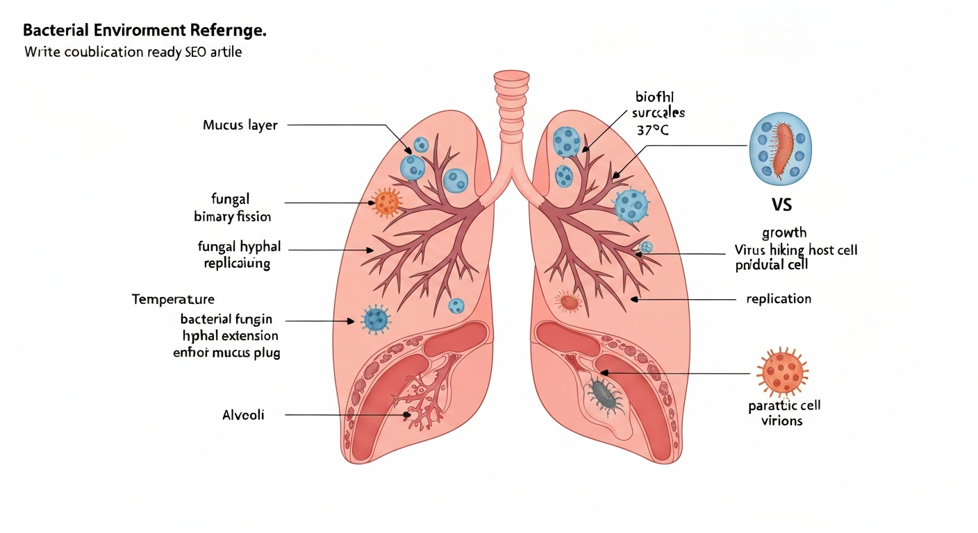
The lung as a microbial environment
To understand which bugs survive and grow in the lung, it helps to think of lung tissue as an environment with specific physical and chemical parameters, the same way food-safety professionals think about water activity, pH, and temperature in a food matrix.
Temperature
Core lung tissue sits at approximately 37°C, right in the ideal growth range for most human pathogens. The upper airways are slightly cooler, and that gradient matters. Some pathogens, like Aspergillus fumigatus, are thermotolerant and can grow comfortably at 37°C and even up to about 50°C, which is part of why they outcompete many environmental molds when they enter the body.
Oxygen gradients
The lung is not uniformly aerobic. Alveoli are well-oxygenated, but deep within mucus plugs, abscesses, and biofilms, oxygen levels drop sharply. This creates microaerobic and anaerobic microenvironments that support different organisms. Obligate anaerobes like oral Bacteroides and Fusobacterium species can survive and grow in these low-oxygen pockets, which is why aspiration pneumonia and lung abscesses often involve these organisms. Pseudomonas aeruginosa is especially notable here: it is primarily aerobic but can shift to anaerobic metabolism within mucus biofilms, which is a key reason it persists so stubbornly in cystic fibrosis airways.
Moisture, mucus, and nutrients
The airway surface is continuously coated with mucus, a gel-like mixture of water, glycoproteins, antimicrobial peptides, immunoglobulins, and cellular debris. For most organisms, this is a nutrient-rich, high-moisture environment. Mucus water activity is high, there is abundant carbon and nitrogen, and the pH of healthy airway surface liquid sits around 6.9 to 7.2. Organisms that can bind to or degrade mucin have a competitive advantage. The surfactant layer in the lower alveoli adds another biochemical complexity: pulmonary surfactant contains lipoproteins that some fungi and bacteria can use as carbon sources.
Biofilms and host immunity
Biofilms are structured communities of microorganisms encased in a self-produced matrix. In the lung, Pseudomonas aeruginosa and Staphylococcus aureus form biofilms that dramatically reduce antibiotic penetration and allow persistence. Host immunity shapes which organisms survive: in an immunocompetent host, most environmental fungi are cleared before they can establish a foothold, but in immunocompromised patients (transplant recipients, people with HIV, those on corticosteroids), organisms like Aspergillus, Cryptococcus, and Pneumocystis jirovecii take advantage of reduced surveillance.
How viruses differ from bacteria, fungi, and parasites
Viruses do not grow in the lung the way bacteria or fungi do. They do not divide by binary fission, they do not consume nutrients from airway secretions, and they cannot be cultured on a standard agar plate. See 'Can viruses grow in culture medium?' (internal resource c159d453-a518-48e7-a7d5-9b2416d5e5db) for more on viral culture requirements and how they differ from bacterial/fungal media. To replicate, a respiratory virus must enter a lung epithelial cell, dismantle the cell's protein-making machinery, and use it to produce copies of itself. This obligate intracellular requirement means environmental factors like oxygen level, pH, or mucus composition have almost no direct bearing on viral replication the way they do for bacterial or fungal growth.
This distinction also affects diagnosis. Bacteria and fungi can be cultured on selective and differential growth media under controlled incubation conditions. Viruses require cell culture lines (living cells that the virus can infect) or, more commonly in modern labs, molecular detection methods like PCR and nucleic acid amplification tests (NAATs). Structure and Classification of Viruses, Medical Microbiology (NCBI Bookshelf) notes that viruses are obligate intracellular parasites and that laboratory diagnosis typically requires cell culture or molecular methods (PCR/NAAT) rather than standard bacteriological culture media Structure and Classification of Viruses — Medical Microbiology (NCBI Bookshelf). The same logic applies when thinking about whether viruses grow in food: they do not replicate in food matrices at all because food lacks living cells with the necessary replication machinery, which is a topic worth understanding separately in environmental food-safety contexts.
Bacteria that grow in the lungs
Here is a practical breakdown of the major bacterial lung pathogens, their preferred niches within the lung environment, and the culture conditions labs use to grow them. Understanding the lab conditions is useful because they reflect the organism's environmental preferences in vivo.
| Organism | Lung niche / disease | Oxygen requirement | Key culture conditions | Notable environmental source |
|---|---|---|---|---|
| Streptococcus pneumoniae | Alveoli, lobar pneumonia (most common community-acquired bacterial pneumonia in the US) | Facultative anaerobe | 5% sheep blood agar, 35–37°C, ~5% CO2 | Human upper respiratory tract (droplet transmission) |
| Staphylococcus aureus | Post-influenza pneumonia, healthcare-associated pneumonia, biofilm-related | Facultative anaerobe | Blood agar, 35–37°C, aerobic or anaerobic | Skin, nares, fomites, healthcare environments |
| Haemophilus influenzae | Chronic obstructive pulmonary disease exacerbations, community pneumonia | Facultative anaerobe | Chocolate agar (hemin + NAD factors), 35–37°C, 5% CO2 | Human respiratory tract (droplet transmission) |
| Pseudomonas aeruginosa | Cystic fibrosis, bronchiectasis, ventilator-associated pneumonia, biofilms in hypoxic mucus | Aerobic, anaerobic in biofilms | Routine blood or MacConkey agar, 37°C | Water, soil, healthcare equipment, drains |
| Legionella pneumophila | Legionnaires' disease (inhalation of aerosols) | Aerobic | BCYE agar (buffered charcoal yeast extract with alpha-ketoglutarate), 35–37°C | Cooling towers, showerheads, hot tubs, water systems |
| Mycobacterium tuberculosis | Pulmonary TB, upper lobe cavitary disease | Obligate aerobe (prefers high O2) | Löwenstein-Jensen, Middlebrook 7H10/7H11, MGIT liquid; BSL-3 required | Aerosols from active TB cases |
| Mycobacterium avium complex / M. abscessus | Chronic pulmonary NTM disease | Aerobic | Mycobacterial media (MGIT, LJ, Middlebrook) after decontamination | Soil, household water, showerheads, plumbing biofilms |
| Nocardia spp. | Pulmonary nocardiosis (immunocompromised hosts) | Obligate aerobe | Routine media, prolonged incubation (days to weeks), modified Kinyoun stain | Soil, decaying vegetation, inhalation |
| Anaerobes (Bacteroides, Prevotella, Fusobacterium) | Aspiration pneumonia, lung abscess | Obligate anaerobes | Anaerobic transport essential; anaerobic chambers or jars, blood/KVLB agar | Oral flora, aspiration of gastric/pharyngeal contents |
| Bordetella pertussis | Whooping cough, tracheobronchitis | Aerobic | Bordet-Gengou or Regan-Lowe agar; PCR preferred in practice | Human respiratory secretions (aerosol/droplet) |
| Mycoplasma pneumoniae | Atypical ('walking') pneumonia | Aerobic (no cell wall) | Specialized SP4/Eaton media, prolonged incubation; PCR/serology preferred | Human respiratory secretions |
A few organisms deserve a closer look because they illustrate how environmental conditions drive pathogen behavior. Mycobacterium tuberculosis prefers the oxygen-rich upper lobes of the lung, which is why classical TB shows upper-lobe infiltrates and cavities. The bacterium grows slowly (doubling time around 15 to 20 hours compared to about 20 minutes for E. coli), which is why TB culture can take weeks. Nontuberculous mycobacteria like M. avium complex and M. abscessus are found in ordinary environmental sources including household plumbing biofilms and showerheads, making them an exposure route with direct relevance to water-safety thinking.
Legionella pneumophila is another organism where environmental control is the primary prevention strategy. It does not spread person to person. Instead, it grows within amoebae in warm water systems (optimally 25 to 45°C) and is released as aerosols from cooling towers, decorative fountains, and inadequately maintained plumbing. Lab recovery uses BCYE agar with alpha-ketoglutarate, and environmental testing of water samples follows CDC-specified concentration and culture protocols.
Fungi that colonize or grow in the lungs
Fungal lung infections are divided into two broad categories: infections caused by environmental fungi that any person might inhale (the endemic dimorphic fungi), and opportunistic infections caused by fungi that only cause disease when host immunity is compromised.
Endemic dimorphic fungi
Histoplasma capsulatum, Coccidioides immitis/posadasii, and Blastomyces dermatitidis are thermally dimorphic, meaning they exist as environmental molds in soil but convert to a yeast form (or in Coccidioides' case, spherules) at body temperature. This temperature-driven morphological switch is a key virulence mechanism. Histoplasma is found in soil enriched with bird or bat droppings and is endemic to the Ohio and Mississippi River valleys in the US. Coccidioides is concentrated in the desert soils of the American Southwest. Inhalation of disturbed soil particles or aerosols is the primary exposure route.
Aspergillus fumigatus
Aspergillus fumigatus is ubiquitous in the environment, its conidia (spores) are present in most outdoor and indoor air samples. Most healthy people inhale conidia daily without consequence because macrophages clear them. In immunocompromised patients, conidia germinate into hyphae that invade lung tissue directly, a condition called invasive pulmonary aspergillosis with high mortality. A. fumigatus is thermotolerant, growing at 37°C and up to approximately 50°C on Sabouraud dextrose agar or inhibitory mold agar. Its thermotolerance at human body temperature is a core reason it outcompetes most other environmental molds as a human pathogen.
Cryptococcus and Pneumocystis
Cryptococcus neoformans is also an environmental organism found in soil and bird droppings (particularly pigeon). After inhalation, it can cause pulmonary cryptococcosis and is capable of spreading to the central nervous system. Pneumocystis jirovecii is different: it is an atypical fungus that cannot be cultured on standard fungal media and is exclusively a human pathogen. It causes Pneumocystis pneumonia (PCP) almost exclusively in severely immunocompromised individuals, particularly those with advanced HIV infection. Its growth parameters are essentially defined by the human lung environment and cannot be reproduced in a standard culture system.
| Fungus | Form in lung | Key growth/culture conditions | Primary at-risk population |
|---|---|---|---|
| Aspergillus fumigatus | Hyphae from germinated conidia | Sabouraud/inhibitory mold agar, 35–37°C; grows up to ~50°C | Immunocompromised (neutropenia, transplant, steroids) |
| Histoplasma capsulatum | Yeast phase at 37°C | Mycobiotic/Sabouraud agar; room temp = mold, 35–37°C = yeast; prolonged incubation | Anyone in endemic areas; occupational/environmental exposure |
| Coccidioides immitis/posadasii | Spherules containing endospores | Sabouraud agar (BSL-3); room temp mold, 37°C spherule conversion | Residents/travelers to US Southwest; field workers |
| Cryptococcus neoformans | Encapsulated yeast | Sabouraud agar with caffeine/canavanine to differentiate; 37°C | HIV/AIDS, transplant recipients |
| Pneumocystis jirovecii | Cysts and trophic forms | Cannot be cultured in vitro; diagnosed by PCR or methenamine silver stain on BAL | Advanced HIV, primary immunodeficiency, high-dose steroids |
| Candida spp. | Yeast (hematogenous spread to lung) | Blood agar, CHROMagar Candida, 35–37°C | ICU patients, central lines, immunocompromised |
For lab identification, temperature manipulation is especially important with dimorphic fungi. A culture grown at room temperature (25 to 28°C) will produce the mold (mycelial) phase, but shifting the same culture to 35 to 37°C triggers conversion to the pathogenic yeast or spherule phase. This thermal conversion is a diagnostic confirmation step, and it mirrors exactly what happens biologically when an inhaled spore encounters the 37°C lung environment.
Parasites with pulmonary stages
True parasites with significant lung involvement are less common in high-income settings but are globally important. The lung is either a permanent niche (for some) or a transient developmental stage (for others).
Paragonimus (lung flukes)
Paragonimus westermani and related species are flatworms (trematodes) that establish themselves permanently in the lung parenchyma, where adult worms live encapsulated in cysts. The transmission route is food-related: humans are infected by eating raw or undercooked freshwater crabs or crayfish containing metacercariae. Once swallowed, the larvae penetrate the gut wall, migrate through the diaphragm, and enter the lung, where they mature into adults, sometimes living for 20 years. For more on the likelihood of parasites developing in food products and related food-safety precautions, see can parasites grow in food. This is a direct food-safety concern, as proper cooking (internal temperature sufficient to kill metacercariae) is the key control point.
Strongyloides and other soil-transmitted helminths
Strongyloides stercoralis undergoes a pulmonary larval migration as part of its normal life cycle. Filariform larvae that penetrate skin travel via the bloodstream to the lung, ascend through the airways, are swallowed, and mature in the intestine. In immunocompromised individuals, hyperinfection syndrome can produce massive larval migration through the lung, causing hemorrhage and secondary bacterial pneumonia. Toxocara species (dog and cat roundworms) cause visceral larva migrans: larvae can migrate through the lung, causing eosinophilic pneumonitis (Löffler syndrome), though the lung is not their terminal destination. These parasites do not replicate in the lung in the conventional sense; rather, they pass through or become encysted.
Filarial larvae and other pulmonary parasites
Tropical pulmonary eosinophilia is caused by an immune reaction to microfilariae of Wuchereria bancrofti and Brugia malayi trapped in the lung capillaries. Echinococcus granulosus (tapeworm) forms hydatid cysts in the lung that can grow to several centimeters in diameter, a genuine slow form of parasitic 'growth' within lung tissue. Diagnosis of pulmonary parasites typically relies on serology, imaging, and direct microscopy or biopsy rather than culture, because most do not have a free-living in vitro growth phase that can be replicated in a laboratory medium.
Culturing lung pathogens: what the lab conditions tell us
The way a lab cultures an organism reveals a great deal about its environmental preferences in the lung. Blood and chocolate agar at 35 to 37°C with CO2 support the fastidious respiratory bacteria that reflect the lung's warm, slightly CO2-enriched microenvironment. Bordetella pertussis is best isolated on selective media (Bordet–Gengou or Regan–Lowe) but in clinical practice is often diagnosed by PCR; see Laboratory Diagnosis of Pertussis, CDC Bordetella pertussis is best isolated on selective media (Bordet–Gengou or Regan–Lowe) but in clinical practice is often diagnosed by PCR; see Laboratory Diagnosis of Pertussis — CDC.. Anaerobic chambers mimic the oxygen-depleted conditions inside lung abscesses. Mycobacterial media with prolonged incubation reflect the slow doubling time of mycobacteria. Fungal media incubated at two temperatures (room temp and 37°C) exploit the thermal dimorphism that drives pathogenicity.
Specimens for lung pathogen recovery include sputum, bronchoalveolar lavage (BAL), bronchial washings, pleural fluid, and lung tissue biopsy. Transport conditions matter enormously. Anaerobes from a suspected lung abscess require anaerobic transport media and fast processing; delay causes false-negative results. Legionella from environmental water sources requires concentration steps before plating on BCYE agar per CDC environmental sampling protocols.
Exposure routes and food-safety relevance
Most lung pathogens reach the airways by one of three routes: inhalation of airborne particles or aerosols, aspiration of oral or gastric contents, or hematogenous spread from another infection site. Food-related transmission is relevant in a few specific cases.
- Paragonimus: direct food-safety issue, raw or undercooked crabs and crayfish carry infective metacercariae. Cooking to safe internal temperature eliminates the risk.
- Legionella: not food-transmitted, but water-system management (temperature control above 60°C or below 20°C, biocide treatment) is the environmental control point, directly parallel to temperature control in food safety.
- NTM (M. avium complex, M. abscessus): linked to household plumbing biofilms and showerhead aerosols, an exposure route that involves water system hygiene rather than food itself.
- Aspergillus and other molds: heavily contaminated grain, compost, or decaying organic matter can serve as high-spore-load aerosol sources, relevant for food-processing facilities and agricultural workers.
- Histoplasma: occupational exposure during construction, demolition, or cleanup of bird/bat-roosting sites, not a food-contact route but relevant to environmental and occupational health risk assessment.
It is worth addressing a misconception that occasionally arises in public health and food-safety conversations: the idea that maggots, mosquito larvae, or worms could grow inside human lungs under normal circumstances. For related information on whether worms can develop in old dog poop, see the article "can worms grow in old dog poop". A related question, can mosquitoes grow in toilet, examines whether mosquito larvae can develop in indoor plumbing, which similarly depends on specific aquatic conditions and is unrelated to lung colonization. For further detail on whether maggots can develop in aquatic environments, see can maggots grow in water. Obligate external parasites and organisms that require specific aquatic or organic substrates simply cannot establish themselves in lung tissue. The lung's immune defenses, temperature, anaerobic microenvironments, and absence of the specific nutrient matrices these organisms need make colonization physiologically impossible in a healthy host. The organisms that genuinely grow in the lung are those specifically adapted to its conditions.
Prevention and control for environmental and food-safety professionals
Prevention of lung infections from environmental and food-related exposures comes down to controlling the source environment, the transmission route, and host susceptibility.
- Water system management: maintain hot water above 60°C and cold water below 20°C to prevent Legionella growth; implement regular flushing, biocide treatment, and monitoring in building water systems, cooling towers, and food-processing equipment that uses water spray.
- Aerosol control in food processing: high-spore environments (grain handling, composting, mushroom production) warrant respiratory protection (N95 or higher) and ventilation controls to limit inhalation of Aspergillus and other thermotolerant mold conidia.
- Cook shellfish thoroughly: ensure freshwater crabs and crayfish reach safe internal temperatures to eliminate Paragonimus metacercariae. This is the single most effective food-safety intervention for lung fluke prevention.
- Soil and occupational exposure controls: workers in demolition, excavation, or sites with bird/bat activity in Histoplasma- or Coccidioides-endemic areas should use appropriate respiratory protection and follow CDC or OSHA guidance for disturbing potentially contaminated soil.
- Healthcare and laboratory biosafety: MTB culture requires BSL-3 containment; Coccidioides in mold phase is also a BSL-3 organism. Lab personnel handling respiratory specimens should use appropriate biosafety levels, proper cabinet work, and PPE.
- Immunocompromised patient precautions: minimize exposure to high-spore environments (construction dust, compost, potting soil) for transplant recipients, patients on long-term corticosteroids, and individuals with HIV/AIDS, as their ability to clear inhaled fungi is significantly reduced.
- Prophylaxis and surveillance: in clinical and occupational contexts, antifungal prophylaxis (for example, fluconazole or posaconazole in high-risk transplant patients) and active environmental monitoring of healthcare facility air quality during construction are established control strategies.
Understanding which organisms can actually grow in the lung, and what environmental conditions enable that growth, is the foundation for rational prevention. The same principles that govern microbial behavior in food and water matrices, temperature, oxygen availability, moisture, pH, nutrient access, and biofilm formation, apply directly to the lung microenvironment. The lung is simply another substrate with its own set of parameters, and the pathogens that thrive there are the ones that have evolved to meet those parameters. For another relevant comparison, see do viruses grow in food.
FAQ
Which microbial organisms can grow or replicate in human lungs (bacteria, fungi, parasites) and how do viruses differ?
Bacteria, fungi (molds and yeasts), and certain parasites (protozoa and helminths) can colonize and replicate in lung tissue or airways under permissive conditions. Examples include Streptococcus pneumoniae, Staphylococcus aureus, Pseudomonas aeruginosa, Legionella pneumophila, Mycobacterium tuberculosis and nontuberculous mycobacteria, Nocardia, Aspergillus spp., Candida spp., Histoplasma/Coccidioides/Blastomyces (dimorphic fungi), and pulmonary parasites (e.g., Strongyloides, Paragonimus). Viruses differ because they are obligate intracellular parasites: they do not grow on culture media or independently in the environment but replicate only inside host cells. Diagnostic approaches therefore differ: viral detection usually uses molecular tests (PCR/NAAT), antigen tests, serology, or cell culture rather than routine bacterial/fungal culture.
What lung-environment parameters permit microbial growth or persistence?
Key parameters that determine growth and persistence in the lungs include: temperature (human core ~37°C; some fungi tolerate higher), oxygen gradients (airways are aerobic but mucus plugs and biofilms create hypoxic/anoxic niches supporting facultative or anaerobic organisms), moisture and mucus (thick mucus provides hydration and nutrients), pH (generally near neutral in airways but can vary in infected sites), nutrient availability (host-derived proteins, iron, surfactant, dead cells), surface availability for adhesion and biofilm formation (epithelium, mucus, foreign devices), and immune status (impaired defenses permit colonization and replication). Microbes adapted to aerosolized dispersal (small spores/conidia, hardy bacteria) reach lower airways more readily.
Which common bacteria grow in lungs and what are their preferred niches and key growth parameters?
- Streptococcus pneumoniae: community-acquired pneumonia; prefers mucosal surfaces, grows best at 35–37°C in CO2-enriched atmosphere; cultured on blood agar. - Staphylococcus aureus: community and healthcare-associated pneumonia, post-influenza; facultative anaerobe, grows at 35–37°C on routine media. - Pseudomonas aeruginosa: opportunist in cystic fibrosis, bronchiectasis, ventilator-associated pneumonia; aerobic but tolerates hypoxic biofilm niches, grows at 37°C on routine media and forms resilient biofilms. - Legionella pneumophila: causes Legionnaires’ disease after inhalation of contaminated aerosols; grows on buffered charcoal yeast extract (BCYE) and prefers aquatic-derived amoeba-associated environments. - Mycobacterium tuberculosis and NTM: slow growers that establish chronic pulmonary infection; require specialized mycobacterial media and can grow at 35–37°C but with prolonged incubation. - Anaerobic oral flora (Bacteroides, Prevotella, Fusobacterium): seen in aspiration pneumonia and lung abscesses; require anaerobic conditions and transport for culture.
Which fungi can grow in the lung and what growth characteristics matter for lab recovery?
Important pulmonary fungi include: Aspergillus fumigatus (invasive aspergillosis; thermotolerant, conidia inhaled from environment; cultured on Sabouraud or inhibitory mold media, incubated ~35°C), Candida spp. (can colonize airways in debilitated patients; grow on routine fungal/bacterial media), dimorphic endemic fungi (Histoplasma, Coccidioides, Blastomyces; inhaled spores convert to yeast in tissue and require prolonged culture with temperature-based phase differentiation), and Cryptococcus neoformans (inhaled yeast, cultured on standard fungal media). Laboratory recovery often uses Sabouraud dextrose or mycobiotic media, selective antibiotics to inhibit bacteria, and longer incubation times; incubation temperature choices (room temp vs 35–37°C) help distinguish mold vs yeast/dimorphic phases.
Which parasites can involve the lung and by what routes do they reach pulmonary tissue?
Parasites that can involve lungs include: - Protozoa: Toxoplasma gondii (dissemination in immunocompromised), Pneumocystis jirovecii (fungal-like organism causing pneumonia in immunosuppressed). - Helminths: Strongyloides stercoralis (larval migration through lungs), Ascaris lumbricoides (larval pulmonary migration), Paragonimus spp. (lung fluke from ingesting undercooked crustaceans). They reach lungs via hematogenous/lymphatic dissemination, larval migratory life cycles, or direct inhalation/aspiration of infectious stages when relevant. Diagnosis uses microscopy, serology, PCR, and sometimes sputum/bronchoalveolar lavage examination.
How are lung pathogens diagnosed and cultured in the laboratory (summary of media and incubation conditions)?
General guidelines: - Routine bacterial respiratory pathogens: blood agar and chocolate agar incubated at 35–37°C; 5% CO2 enhances growth of fastidious respiratory bacteria (e.g., Streptococcus pneumoniae, Haemophilus influenzae). - Fastidious organisms: Haemophilus requires X/V factors or chocolate agar; Bordetella pertussis uses Bordet–Gengou or Regan–Lowe medium (culture often replaced by PCR). - Legionella: buffered charcoal yeast extract (BCYE) agar with alpha‑ketoglutarate; culture of lower respiratory secretions or environmental water concentrates. - Mycobacteria: Löwenstein–Jensen, Middlebrook 7H10/7H11, or liquid MGIT systems with prolonged incubation under BSL‑3 precautions for MTB. - Anaerobes: require anaerobic transport and incubation in anaerobic chambers or jars. - Fungi: Sabouraud dextrose or inhibitory mold media; incubate at appropriate temperatures (room temp and 35°C) and allow prolonged incubation for dimorphic fungi. - Viruses: detected by NAAT/PCR, antigen tests, or cell culture (specialized) rather than routine bacterial/fungal media. Clinical labs choose tests based on suspected pathogens and specimen type (sputum, BAL, pleural fluid, nasopharyngeal swab).
Next Article
Can Parasites Grow in Food? Risks, Timing, and Safety Tips
Learn if parasites can develop after food is stored, plus cooking, freezing and handling tips to reduce infection risk.