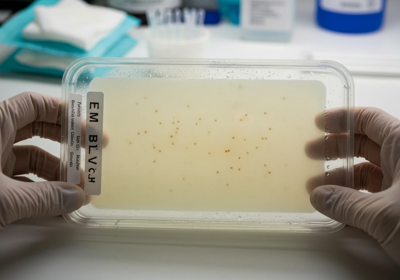
Staphylococcus aureus and Staphylococcus epidermidis do not grow reliably on EMB (eosin methylene blue) agar. Both are gram-positive, and EMB is specifically designed to inhibit gram-positive organisms while selecting for gram-negative enteric bacteria. If you plate either Staphylococcus species on EMB, expect little to no recovery. The agar's dye combination (eosin Y and methylene blue) does the inhibiting, and the selective pressure is real enough that HiMedia's quality control data lists S. aureus ATCC 6538 as showing 0% recovery on Levine EMB under standard QC conditions.
Does Staphylococcus aureus Grow on EMB Agar? Plate Results

Marcus Reeves
6 Jun 2026
The direct answer: S. aureus and S. epidermidis on EMB
Neither species is expected to grow on EMB agar under normal lab conditions. S. aureus is inhibited, and S. epidermidis fares no better. Both are gram-positive cocci, and EMB's dyes (eosin Y at 0.04 g/L and methylene blue at 0.065 g/L) are bacteriostatic toward gram-positive cell walls at the pH range the medium maintains (around 7.1 ± 0.2). This is not a quirk of one brand or formulation, it is how EMB is supposed to work, and it is consistent across Levine-formulation products from Thermo Fisher, HiMedia, and others.
That said, Thermo Fisher's own IFU notes that "organisms other than gram-negative enteric bacilli can grow on EMB agar", meaning breakthrough growth is possible in some conditions, particularly with heavy inocula or certain strain variations. If you do see colonies, do not assume it means you have a coliform. It more likely means the inhibition was incomplete, and you need confirmatory testing before drawing any conclusions.
How EMB works, and why it does not suit Staphylococcus
blank" rel="noopener noreferrer">EMB agar has two jobs: it selects against gram-positives, and it differentiates among gram-negatives based on lactose fermentation. The lactose (10 g/L in the Levine formulation) provides the fermentable sugar. When a bacterium ferments it quickly and drops the local pH below roughly 5, eosin Y and methylene blue form a complex that precipitates onto the colony surface. That precipitation is what creates the striking blue-black color with a green metallic sheen you see with rapid lactose fermenters like E. coli. Slower or non-lactose fermenters produce little or no acid locally and show up as colorless or faintly pink colonies. Gram-positive organisms, including both Staphylococcus species, never get far enough in the growth cycle to show any of this because the dyes suppress them before meaningful colony formation occurs.
Plates are read after incubation at 35 to 37°C for 24 to 48 hours. By 24 hours, the differential patterns for true gram-negative targets are usually clear. If you are not seeing those patterns, and especially if you are trying to identify a Staphylococcus isolate, EMB is simply the wrong plate for the job. MSA (mannitol salt agar) is designed to recover and differentiate Staphylococcus, so it is not a reliable medium for growing gram-negative bacteria. For staphylococci, MSA (mannitol salt agar) is a better choice than EMB because it is selective for them and can differentiate S. epidermidis from mannitol-fermenters does S. epidermidis grow on MSA plate.
Growth on blood agar vs EMB vs nutrient agar, a quick comparison
If you are trying to understand where Staphylococcus species will actually grow, the medium choice matters enormously. Here is how the three common media compare for these organisms:
| Medium | S. aureus growth | S. epidermidis growth | Key observation |
|---|---|---|---|
| EMB agar (Levine) | Inhibited / 0% QC recovery | Inhibited | Dyes suppress gram-positives; not appropriate for Staphylococcus |
| Blood agar (sheep) | Good growth; beta-hemolysis (clear zone around colonies) | Good growth; non-hemolytic (gamma) | Hemolysis pattern is a key differentiator between the two species |
| Nutrient agar | Good growth; creamy, opaque colonies | Good growth; smaller white colonies | Non-selective; no differentiation between species or gram class |
Blood agar is far more useful when working with Staphylococcus. S. aureus produces large, creamy white to golden colonies with beta-hemolysis, a complete clearing of red blood cells around the colony. S. epidermidis produces smaller, white, raised, cohesive colonies about 1 to 2 mm after overnight incubation, with no hemolysis at all. That hemolysis difference is one of the first practical clues you get at the plate level. Nutrient agar grows both species fine but gives you nothing to differentiate on. EMB gives you inhibition and nothing else.
What colony morphology looks like when you see it on EMB (and why it can mislead you)
If Staphylococcus does break through on EMB, which can happen with a very heavy inoculum, the colonies will not look like typical coliform colonies. You will not see the dark, metallic green sheen characteristic of E. coli-type lactose fermenters, because that sheen requires rapid acid production from lactose fermentation. Staphylococci do not ferment lactose in the same way, so the dye complex does not precipitate the same way. Any breakthrough colonies tend to be pale, small, and non-shiny. They can also look colorless or faintly pigmented depending on the strain.
S. aureus strains may or may not produce their characteristic golden-yellow pigment depending on conditions, so do not count on color alone even on better-suited media. On EMB specifically, any pigment is likely masked or absent. The absence of metallic sheen is not enough to call a colony Staphylococcus, plenty of gram-negative non-lactose fermenters also appear colorless on EMB. This is exactly why EMB results for any suspected Staphylococcus isolate need follow-up.
Confirming your isolate, practical next steps
If your goal is to confirm whether you are dealing with S. aureus or S. epidermidis, move off EMB and use media and tests that are actually designed for staphylococci. Here is a practical sequence:
- Gram stain first: Staphylococci are gram-positive cocci in clusters. If your EMB isolate is gram-negative, stop here — it is not Staphylococcus.
- Catalase test: Staphylococci are catalase-positive (bubbling with 3% H2O2). Streptococci are catalase-negative. This quickly rules out strep contamination.
- Coagulase test: S. aureus is coagulase-positive. S. epidermidis is coagulase-negative. The slide test (clumping factor) is fast; a tube coagulase test at 4 hours is the gold standard. Latex agglutination kits (which detect clumping factor and protein A) are widely used and correlate well with tube coagulase results.
- Plate on blood agar and observe hemolysis: S. aureus produces beta-hemolysis; S. epidermidis produces none. This step alone separates most clinical and food lab cases.
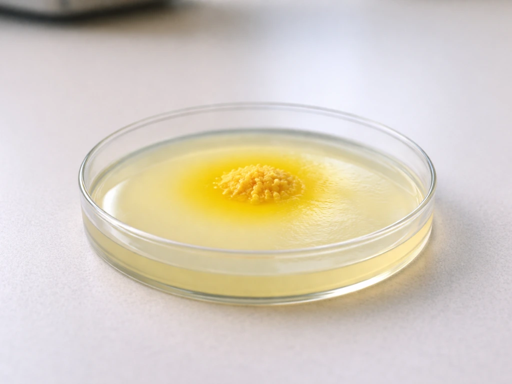
- Consider mannitol salt agar (MSA) if you want a selective confirmation: S. aureus ferments mannitol and produces yellow colonies with yellow halos from the pH indicator change. Coagulase-negative staphylococci like S. epidermidis grow on MSA but produce pink or red colonies without the yellow zone.
- For MRSA confirmation: if methicillin resistance is clinically relevant, PBP2a latex agglutination or immunochromatographic assay (or mecA PCR) is appropriate.
The catalase-plus-coagulase combination is the workhorse here. It is fast, cheap, and gives you a presumptive identification of S. aureus in under an hour in most lab settings. Latex agglutination adds speed and reliability when available. Relying on EMB colony appearance to make any identification call about Staphylococcus is not sound practice, the medium is not designed for it, and the results are not interpretable in the same framework you would use for enteric gram-negatives.
What this means for food safety and contamination work
EMB (particularly Levine's formulation) is the FDA BAM-referenced medium for coliform recovery in food testing, and it works well for that purpose. If you are running food safety screening and you are looking for coliforms or E. coli, EMB is appropriate. If you are trying to detect or confirm Staphylococcus contamination, for example in a ready-to-eat food, dairy product, or environmental swab, EMB is the wrong tool. Using it as a screening medium for S. aureus will produce false negatives or uninformative results. Mannitol salt agar is the standard selective/differential choice for staphylococci in food contexts, and it gives you actual interpretable results: S. Micrococcus luteus is generally not expected to grow and ferment mannitol on mannitol salt agar under standard conditions Mannitol salt agar is the standard selective/differential choice for staphylococci in food contexts. aureus ferments mannitol and turns the indicator yellow, while other staphylococci do not. That interpretive clarity is what makes medium selection matter so much in practical lab and food safety work.
FAQ
If I see tiny colonies on EMB after plating a Staphylococcus isolate, does that mean S. aureus is growing normally?
Not necessarily. Breakthrough growth can occur with very heavy inocula or if the medium or incubation conditions deviate, and those colonies are often pale and non-shiny rather than matching typical coliform appearance. Any suspect Staphylococcus colonies should be confirmed with staphylococcus-specific follow-up tests, such as catalase and coagulase (or an equivalent identification workflow).
What EMB colony appearance would suggest gram-negative coliforms rather than Staphylococcus breakthrough?
Look for a dark colony with a metallic green sheen, which is linked to rapid lactose fermentation and acid precipitation on EMB. If colonies are colorless, faintly pink, or lack sheen, they are not automatically Staphylococcus either, since non-lactose fermenting gram-negatives can appear similar and still require confirmation.
Does storage time or dehydration of EMB plates change whether Staphylococcus grows?
Yes. If plates dry out, warm up excessively, or are held too long before use, the selective and differential performance can shift. That can increase the chance of rare breakthrough or poor differentiation, so use plates within the manufacturer’s stated window, store them properly, and keep incubation conditions consistent.
Can incubation time (24 versus 48 hours) make Staphylococcus look like it grew on EMB?
Longer incubation can sometimes allow delayed growth of low numbers of organisms to become visible, but it still does not make EMB a reliable staphylococcal medium. If you are comparing plates, keep the incubation window consistent and rely on confirmatory testing rather than time-dependent interpretation.
Is Levine EMB the same as other EMB agar brands for deciding whether S. aureus grows?
The general behavior is similar across Levine-formulation products, since the key dyes and intended pH range drive inhibition of gram-positives. However, small differences in formulation, dye concentration, or QC performance can affect how often breakthrough colonies appear, so brand consistency and plate QC still matter if you routinely get unexpected results.
What is the right alternative plate if my purpose is to check for S. aureus or S. epidermidis?
Use mannitol salt agar (MSA) or another staphylococcus-selective medium for recovery and interpretation. MSA is selective for staphylococci and the mannitol indicator gives actionable differentiation, especially distinguishing mannitol fermenters like S. aureus from non-fermenters such as many strains of S. epidermidis.
If EMB is for coliforms, how should I confirm suspected Staphylococcus contamination from an EMB plate?
Treat EMB results as non-interpretable for staphylococci. Subculture any suspicious colonies onto blood agar or MSA, then run staphylococcus-oriented tests (for example catalase plus coagulase, or a validated identification system). This avoids false negatives and prevents mislabeling gram-negative organisms as Staphylococcus.
Does Staphylococcus pigment affect interpretation on EMB?
Usually not in a reliable way. Even when S. aureus can produce a golden-yellow pigment on better-suited media, the EMB dye system can mask or suppress visible pigment, so color alone on EMB should never be used to confirm species identity.
Next Article
Does S. epidermidis Grow on MSA Plates and How to Tell
Find out if Staphylococcus epidermidis grows on MSA, typical mannitol color change, and why Streptococcus usually won’t.