Most gram-negative bacteria will not grow on mannitol salt agar (MSA) under standard lab conditions. The 7.5% NaCl in the medium kills or inhibits the vast majority of them before they can form colonies. That said, "gram-negative" is not an automatic disqualifier. A small group of naturally salt-tolerant gram-negative organisms, including some halophiles like Halomonas and occasionally strains of Aeromonas, can survive and even form colonies on MSA. So if you are seeing unexpected growth on an MSA plate, the gram-stain alone does not tell you whether the result is real or a contamination problem. The salt tolerance of the specific organism is what actually matters.
Can Gram-Negative Grow on MSA? What to Expect and Why

Marcus Reeves
23 Jun 2026
What MSA actually is and why it works the way it does

Mannitol salt agar is both a selective and differential medium. The FDA BAM formula (M97) specifies 75 g/L NaCl, 10 g/L mannitol, 0.025 g phenol red, and a final pH of 7.4 ± 0.2, autoclaved at 121°C for 15 minutes. That 75 g/L NaCl works out to roughly 7.5%, which creates a high-osmotic-stress environment that wipes out most non-halotolerant bacteria regardless of their gram stain.
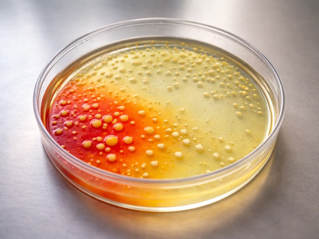
The differential part comes from the mannitol and phenol red combination. Phenol red has a pK of around 7.8, so it sits in the red-to-yellow transition zone at physiological pH. When an organism ferments mannitol, it produces acidic end-products that drop the pH and shift the indicator from red to yellow, either in the colonies themselves or as a halo in the surrounding agar. Organisms that grow but do not ferment mannitol leave the agar red or produce colorless colonies against a red background.
The intended purpose of MSA in food safety and clinical microbiology is isolation and presumptive identification of Staphylococcus aureus. Merck’s technical data sheet describes mannitol salt phenol-red agar as a medium for the blank" rel="noopener noreferrer">isolation and presumptive identification of Staphylococcus aureus in food and related contexts, with yellow colonies indicating mannitol fermentation. It is used in food testing workflows (FDA BAM, Merck, Remel/Thermo) specifically because S. aureus is halotolerant and a mannitol fermenter, giving it a clear signature: growth plus yellow colonies. Related organisms like Staphylococcus epidermidis also grow on MSA but do not ferment mannitol, so they produce colorless or pale colonies on a red background.
Can gram-negative organisms actually grow on MSA?
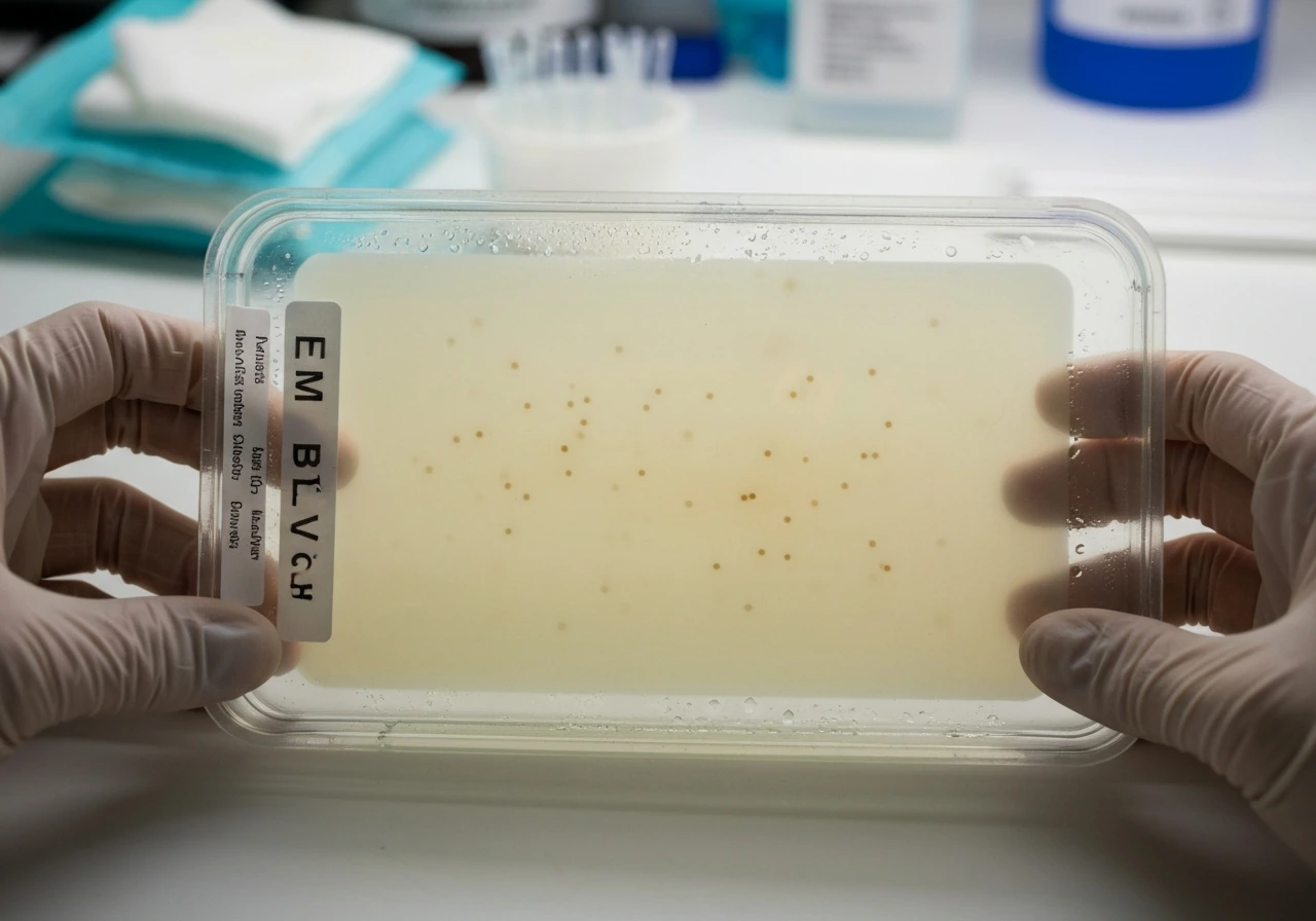
Under standard incubation at 35 to 37°C for 18 to 24 hours, most gram-negative bacteria are inhibited on MSA. E. coli is the textbook example: ASM's own MSA protocol uses E. coli as a control organism specifically to demonstrate that it should not grow. If you see robust E. coli-like colonies on MSA, that is a red flag for a media or contamination problem.
The exceptions are organisms that have evolved to tolerate high salt concentrations. Halomonas, a gram-negative halophilic genus, can form colonies on high-NaCl solid media and would not be suppressed the way E. coli would. Some Aeromonas strains have also shown growth at NaCl concentrations approaching the range MSA provides, depending on the strain. These are not common food-safety pathogens you would expect to encounter in routine testing, but they are real biological exceptions to the "gram-negatives don't grow on MSA" rule.
The practical takeaway: if you are doing routine food safety or clinical microbiology and you see unexpected growth on MSA, your first assumption should not be that you have found a salt-tolerant gram-negative organism. Your first assumption should be contamination, satellite colonies from a nearby heavy growth area, or a media preparation issue. Genuine halophilic gram-negative growth is the explanation of last resort, not the first.
How the selective agents in MSA affect gram-negative bacteria
The 7.5% NaCl is the main gatekeeper. At that concentration, osmotic pressure exceeds what most gram-negative cell walls and membranes can manage. Gram-negative bacteria have an outer membrane that does provide some protection against certain agents, but it does not confer halotolerance. Salt tolerance is a physiological trait tied to osmotic regulation mechanisms, not gram-stain category.
Because the selectivity is entirely osmotic rather than based on something like bile salts or antibiotics, any organism with sufficient halotolerance, regardless of gram-stain result, can potentially survive. This is why VUMicro and other sources correctly point out that MSA is selective for halotolerant organisms, not exclusively for gram-positives. It happens that most halotolerant human-relevant pathogens are gram-positive staphylococci, which is why the medium is described in shorthand as "selective for gram-positives."
The mannitol and phenol red components have no selective function against gram-negative organisms. They only reveal fermentation behavior in organisms that already survived the salt barrier. So if a salt-tolerant gram-negative organism does grow, the yellow-vs-red color reading still tells you whether it fermented mannitol, which is useful differential information but does not identify it as S. aureus.
Interpreting what you see on the plate: growth, color, and what they mean
The two variables to read are presence/absence of growth and whether the phenol red indicator has shifted. Here is how to think through each combination:
| What you see on the plate | Most likely explanation | Next step |
|---|---|---|
| No growth | Organism inhibited by 7.5% NaCl (typical for gram-negatives and many gram-positives) | Expected result for non-halotolerant organisms; no further work needed unless controls also failed |
| Growth, yellow colonies or yellow halo | Halotolerant organism that ferments mannitol; classic S. aureus result | Confirm with coagulase test or other S. aureus confirmatory assay; yellow alone does not equal S. aureus |
| Growth, red or colorless colonies | Halotolerant organism that does NOT ferment mannitol (e.g., S. epidermidis, some other coagulase-negative staph) | Gram stain + additional biochemical tests to identify |
| Sparse or pinpoint growth | Partial inhibition, satellite effect, or very low inoculum surviving on trace nutrients | Check inoculum load, streaking technique, and rule out contamination |
| Unexpected yellow in uninoculated areas or background | Contamination, mixed culture, or media preparation issue (degraded phenol red or incorrect pH) | Check negative control plates, retest with fresh media |
One thing worth stressing: yellowing on MSA tells you that mannitol was fermented, not that the organism is S. does staphylococcus aureus grow on emb agar is a closely related question to ask when you see yellow or robust colonies on MSA yellowing on MSA. aureus. A salt-tolerant gram-negative that ferments mannitol would also produce blank" rel="noopener noreferrer">yellow colonies. This is why confirmatory testing (coagulase, DNase, MALDI-TOF, or similar) is required before calling a yellow MSA colony S. aureus in a food safety context. The color just gets you to the next test, not to a final answer.
Conversely, if an organism fails to grow on MSA, you cannot conclude it is mannitol-negative. As VUMicro correctly notes, growth failure on MSA only means the organism could not tolerate the salt. Its fermentation behavior is simply untestable on this medium.
Troubleshooting when your MSA results do not look right
MSA plates that give you unexpected results, whether that is no growth where you expected some, growth where you expected none, or wrong colony colors, are usually pointing to one of a small number of fixable problems. Work through these in order before re-interpreting the biology.
Check your media preparation first
NaCl concentration is everything on MSA. If your weigh-out or dilution was off, the selective pressure changes dramatically. A batch prepared at 5% NaCl instead of 7.5% will allow gram-negative organisms that would otherwise be inhibited to grow. Always verify the lot and confirm autoclave conditions: 121°C for 15 minutes per FDA BAM M97. Over-autoclaving can degrade phenol red and give you plates that will not shift color correctly even when mannitol is fermented. A Reddit discussion documented exactly this kind of failure, where plates stayed red despite confirmed S. aureus inoculation, pointing to degraded indicator.
Verify your incubation conditions
Standard incubation for MSA is 35 to 37°C for 18 to 24 hours. Himedia's technical document and FDA BAM confirmatory steps both use this window. Incubating at lower temperatures can slow or prevent growth even for halotolerant organisms and delay the phenol red shift. Extended incubation beyond 48 hours can sometimes allow marginally tolerant organisms, including some gram-negatives, to form small colonies as the medium ages, which can create confusion. Read plates at the recommended time.
Confirm your organism's identity and viability
If you are testing whether a specific organism grows on MSA, make sure your stock culture is correctly identified and viable. If you are trying to determine where Staphylococcus aureus grows best, it helps to consider both its preferred growth temperature and the nutrients and salt levels in the environment If you are testing whether a specific organism grows on MSA. Use ATCC-recommended QC strains: S. aureus ATCC and S.
epidermidis ATCC are the standard positive and negative controls listed in Remel's IFU. Running E. coli as a negative control (expected: no growth) is also standard practice per ASM's MSA protocol. If your positive or negative controls are not performing as expected, the problem is the media or the organism, not the biology of gram-stain selectivity.
Look at your streaking technique and inoculum load
Too heavy an inoculum can lead to satellite growth around the main streak, where cells that are not actually tolerating the salt are surviving in nutrient-rich zones created by neighboring lysed or growing cells. This can look like unexpected gram-negative growth. Streak for isolation so that individual colonies are well separated, and use a moderate inoculum rather than a heavy one.
Rule out contamination before assuming unusual biology
Unexpected yellow colonies or unusual growth patterns on MSA should always prompt a contamination check. A mixed culture or an environmental contaminant introduced during plating can produce misleading results. Gram-stain any unexpected colonies directly. If the result is gram-negative, repeat the test from a fresh pure culture before drawing any conclusions about the organism's salt tolerance. As one Reddit discussion noted about erroneous yellowing on MSA, mixed cultures and environmental contamination are the most common explanation for results that do not fit the expected pattern. As another Reddit discussion about erroneous yellowing on MSA points out, mixed cultures and environmental contamination are the most common explanation for results that do not fit the expected pattern.
The bottom line for your MSA work
Gram-negative bacteria as a group do not grow on MSA under standard conditions, but that is because most gram-negatives are not salt-tolerant, not because of anything specific to the gram-negative cell wall. Salt-tolerant gram-negative exceptions do exist in nature. In routine food safety or clinical microbiology, unexpected gram-negative growth on MSA almost always means a media, technique, or contamination problem rather than a genuine halophilic organism.
Micrococcus luteus is sometimes discussed in relation to whether it can grow on mannitol salt agar, so results depend on the strain and its salt and mannitol tolerance. Use your controls consistently, verify your media prep, and always follow up any MSA presumptive result with a confirmatory test before making a final identification call.
FAQ
I see gram-negative colonies on MSA. How can I tell if they are true growth or satellite colonies?
If you see growth, first confirm you do not have a mixed culture. Restreak for isolation from the original sample, then perform a gram stain and a confirmatory ID on colonies chosen from single, well-separated colonies. Salt-tolerant halophiles exist, but contamination and satellite colonies are far more common explanations for unexpected gram-negative-looking growth.
Does yellow color on MSA automatically mean the organism is Staphylococcus aureus?
No, you cannot treat an MSA phenol red color as an identity test for Staphylococcus aureus. Yellowing only indicates mannitol fermentation among organisms that survived the salt. You still need a confirmatory test such as coagulase and/or MALDI-TOF, especially when the colony’s Gram reaction is not what you expected.
If something doesn’t grow on MSA, can I conclude it is mannitol-negative?
If a culture does not grow on MSA, it does not prove it is non-mannitol fermenting. MSA’s 7.5% NaCl creates a selective barrier, so fermentation of mannitol cannot occur unless the organism can tolerate the salt. Use an appropriate non-selective mannitol medium or an alternative assay to test mannitol metabolism.
What should I do if my QC organisms do not behave as expected on MSA?
Use a strict workflow: check media lot preparation records, then verify sterility and indicator performance with the expected QC strains (S. aureus as a positive, S. epidermidis as a presumptive negative, and E. coli as a negative control for growth). If controls fail, troubleshoot preparation, autoclave impact on phenol red, and incubation conditions before interpreting any unknowns.
Is it okay to read MSA at 48 hours or later if colonies are small?
If you incubate longer than the recommended window, small colonies can appear from marginally tolerant contaminants or organisms that otherwise would not establish growth. Read and document results at 18 to 24 hours under the specified temperature range, then if needed, record what changes by a second time point rather than calling the plate based on late growth alone.
How do incubation temperature and timing affect MSA color and colony appearance?
Yes, mismatched incubation conditions can bias results. Incubating colder can delay both growth and the phenol red shift, and using warmer conditions can speed growth for salt-tolerant organisms or increase the chance of confusing artifacts. Follow the standard incubation temperature and timing, and if you deviate, document it because interpretation thresholds may no longer apply.
Could inoculum size or streaking technique cause misleading growth on MSA?
Heavy inoculum and poor streak technique can create a nutrient-rich microenvironment, enabling cells that cannot truly tolerate the salt to survive near lysed neighbors. Use a moderate inoculum, streak for well-isolated colonies, and if you see spreading or lots of tiny satellite colonies, repeat from a purer, isolated colony.
What are good next steps if I really think I have a halotolerant gram-negative on MSA?
When you suspect a halotolerant gram-negative, the best next step is confirmatory identification from a pure colony, not additional reliance on MSA color. Consider testing in a different medium that does not have the same salt barrier if you need to separate salt tolerance from mannitol fermentation, and always re-verify with fresh plates.
What could cause MSA plates to stay red even with Staphylococcus aureus controls?
Phenol red indicator failure can lead to “no color change” even when mannitol fermentation occurs. If plates stay red despite confirmed S. aureus inoculation, suspect degraded indicator from improper autoclaving, overheating, or storage issues, then retest with fresh media and QC strains.
How sensitive is MSA to small errors in NaCl concentration during preparation?
Variations in NaCl concentration are a common root cause of unexpected gram-negative growth. Even modest deviations can change the selective pressure enough to permit organisms that should be inhibited, so verify weigh-outs or pre-poured lot specifications, confirm pH and preparation steps per your protocol, and compare with QC performance.
Next Article
Does Staphylococcus aureus Grow on EMB Agar? Plate Results
See if Staphylococcus aureus grows on EMB agar, what colonies look like, and how to confirm beyond EMB.