Yes, Staphylococcus epidermidis grows on Mannitol Salt Agar (MSA). It tolerates the high salt concentration that kills off most other bacteria, so you will see actual colony growth on the plate. The key thing to know is that it does not ferment mannitol, so the phenol red indicator in the agar stays its original red or pinkish color around the colonies. No yellow halo means no mannitol fermentation, which is exactly what you expect from S. epidermidis and is how you tell it apart from Staphylococcus aureus.
Does S. epidermidis Grow on MSA Plates and How to Tell

Marcus Reeves
22 May 2026
What MSA actually does: salt selection and mannitol differentiation
Mannitol Salt Agar is a two-job medium. Its first job is selection: it contains 75 g of sodium chloride per liter, which works out to about 7.5% NaCl. That concentration is lethal or severely inhibitory to most non-staphylococcal bacteria, including gram-negatives and streptococci. Gram-negative bacteria generally cannot grow on MSA because the high salt concentration is too inhibitory, unless they are unusually salt-tolerant can gram-negative grow on MSA. Only halotolerant organisms, mainly staphylococci, survive and form visible colonies.
Its second job is differentiation: the medium also contains 10 g of mannitol per liter and 0.025 g of phenol red as a pH indicator, with a target pH of 7.4. When a bacterium ferments mannitol, it produces acid, which drops the local pH and blank" rel="noopener noreferrer">turns the phenol red indicator yellow around those colonies. If there is no mannitol fermentation, the indicator stays red or pink. blank" rel="noopener noreferrer">The FDA BAM formulation (M97) and the standard Chapman formula used by manufacturers like Hardy Diagnostics match these numbers exactly, so you can trust this behavior across most commercial lots.
Does S. epidermidis grow on MSA?
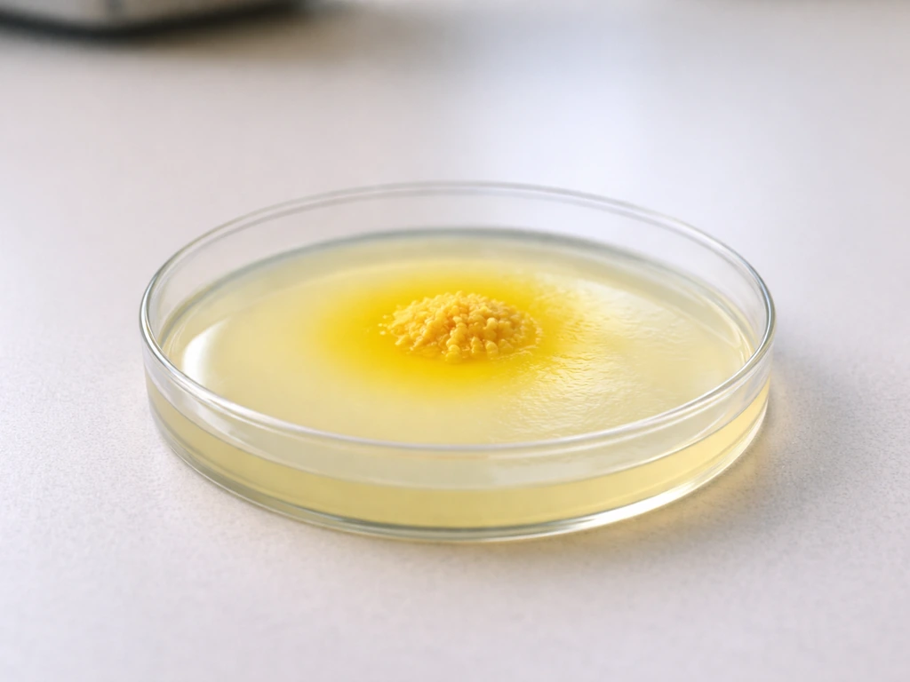
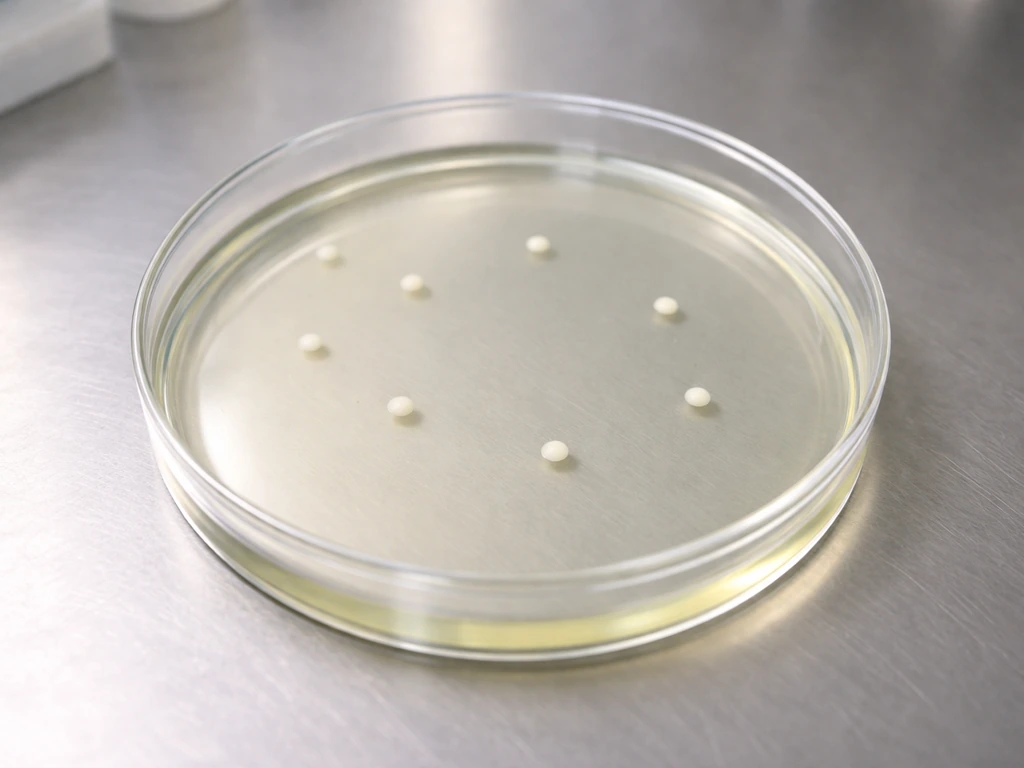
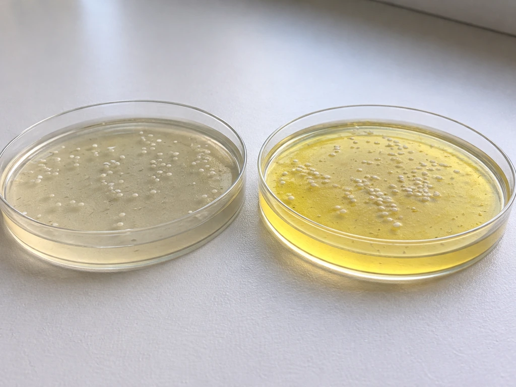
It does. S. epidermidis is halotolerant, meaning it can handle high-salt environments, so the 7.5% NaCl that is supposed to inhibit non-staphylococci does not stop it. On a properly prepared plate incubated at 37°C for 24 to 48 hours, you will typically see small to medium white or off-white colonies, sometimes with a slightly glossy or buttery appearance. The colonies are usually smaller than the glossy gold or yellow-pigmented colonies of S. aureus.
What S. epidermidis does not do is ferment mannitol under standard conditions. It lacks the enzymatic pathway to acidify mannitol efficiently, so the phenol red in the surrounding agar remains unchanged. This is the classic distinguishing result: growth present, color change absent.
What the colonies and agar look like: what to expect when you pull the plate
When you read an MSA plate inoculated with S. epidermidis, here is what you are looking at in practice:
- Colony color: white to off-white or pale cream, non-pigmented
- Colony size: typically 1 to 2 mm after 24 to 48 hours at 37°C, sometimes slightly larger with extended incubation
- Colony texture: smooth, slightly raised, and opaque
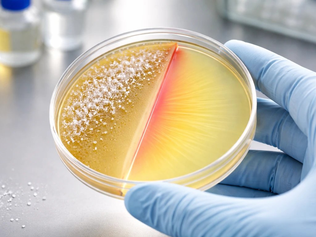
- Agar color around colonies: red or pink (unchanged phenol red), no yellow halo
- No zone of acidification in the surrounding medium
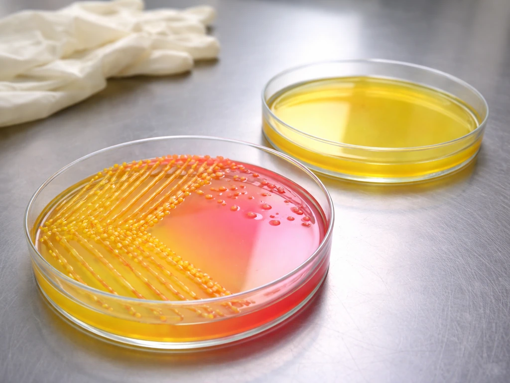
Compare this directly to S. aureus, which produces a bright yellow halo in the agar from mannitol fermentation and often shows golden or yellow-pigmented colonies. If you are distinguishing S. aureus from similar staphylococci on MSA, it can also help to know where does staphylococcus aureus grow in the first place.
S. aureus can also grow on MSA, but it is recognized by a bright yellow halo from mannitol fermentation. That yellow color change is the single most useful visual cue on the plate. If you have growth but no yellow, your first suspect is a mannitol-negative staphylococcus like S.
epidermidis. If you have growth and yellow all around those colonies, lean toward S. aureus until you can confirm with additional tests.
Can Streptococcus grow on MSA?
Under standard conditions, no. Streptococcus species are not halotolerant the way staphylococci are. The 7.5% NaCl in MSA inhibits streptococcal growth effectively, which is exactly the point of the salt selection. You should not see streptococcal growth on a properly prepared, correctly incubated MSA plate. Does Micrococcus luteus grow on mannitol salt agar? It depends on its salt tolerance and ability to ferment mannitol.
The confusion usually comes from two places. First, some people conflate Staphylococcus and Streptococcus, especially in early coursework, and expect similar behavior on selective media. They are different genera with different salt tolerance. Second, enterococci (which were once classified as streptococci and are still casually grouped with them by some) are more salt-tolerant and can grow on MSA, which can be misleading if you are not expecting it. True Streptococcus pyogenes, Streptococcus pneumoniae, and similar pathogens will not form visible colonies on MSA.
If you are working with a mixed culture and see unexpected growth, do not assume streptococcal contamination is the problem. Check whether the isolate is a staphylococcus that you did not expect, or consider enterococcal contamination if your sample source makes that plausible.
How to read your plate: growth vs color change
Reading an MSA plate correctly means treating growth and color change as two separate, independent observations. Ask yourself both questions every time you read a plate.
| What you see on MSA | Most likely organism | What it means |
|---|---|---|
| Growth + yellow halo in agar | S. aureus (presumptive) | Halotolerant AND mannitol-fermenting; run confirmatory coagulase test |
| Growth + no color change (red/pink agar) | S. epidermidis or other coagulase-negative staph | Halotolerant, mannitol-negative; not presumptively S. aureus |
| No growth | Non-staphylococcal organism (e.g., Streptococcus, gram-negatives) | Inhibited by salt; organism is not halotolerant |
| Faint or pinpoint growth + no color change | Possible non-staphylococcal contaminant or stressed cells | Recheck incubation conditions and medium; consider restreaking |
| Growth + faint yellow only near center of heavy growth | Possibly S. epidermidis with high inoculum, or mixed culture | Do not call as S. aureus on color alone; isolate and confirm |
The last row in that table is worth emphasizing. A very heavy inoculum of S. epidermidis can sometimes produce a slight local pH shift that looks like faint yellowing, especially in the dense center of a streak. Do not call that a positive mannitol result. True S. aureus fermentation produces a clear, bright yellow halo extending well into the surrounding agar, not just a subtle tint under a thick colony mass.
Troubleshooting your MSA plate result
If your result does not match what you expected, work through these checks before drawing conclusions.
Check the medium itself
- Confirm the agar is MSA and not a different selective medium (label mix-ups happen more often than people admit)
- Verify the agar was stored at 2 to 8°C and not frozen or exposed to prolonged light, which can degrade phenol red
- Check the lot expiration date; old plates can lose differential ability even if the agar looks fine
- Plates should be pre-warmed to 35 to 37°C for at least 30 minutes before inoculation to avoid condensation and cold-shock inhibition
Check incubation conditions
- Incubate at 35 to 37°C for a full 24 to 48 hours before reading; reading at 18 to 20 hours may miss slow growers
- Confirm the incubator is actually holding temperature with a calibrated thermometer, not just the display readout
- Do not stack plates more than two or three high; uneven heat distribution affects growth uniformly but can cause patchy color development
- Plates incubated beyond 48 hours can show false color shifts from metabolic byproducts unrelated to mannitol fermentation
Check your inoculum
- Use a pure culture; mixed inocula are the most common reason for confusing results on MSA
- Avoid an inoculum that is too heavy; overloading the plate can suppress the indicator reaction or create false acid zones
- If working from a broth culture, ensure the broth was not overgrown or contaminated before plating
- If you are testing an unknown from a patient or food sample directly, expect mixed flora and plan to sub-culture isolated colonies before making any final call
Check your interpretation logic
- Growth alone on MSA does not identify the organism to species; it only tells you it is halotolerant
- Mannitol fermentation (yellow) is a presumptive positive for S. aureus, but always confirm with a coagulase test before reporting
- No color change with growth is consistent with S. epidermidis and other coagulase-negative staphylococci, but is not proof of identity
- No growth does not rule out staphylococci if the medium or incubation was compromised; run a control streak of a known S. aureus or S. epidermidis strain alongside unknowns if results seem off
MSA is a reliable, well-characterized medium when it is handled correctly. The selectivity for halotolerant staphylococci and the mannitol color differentiation between S. aureus and S. epidermidis have been consistent across decades of use and across multiple standard formulations. If your result looks wrong, the answer is almost always in the checklist above rather than in unexpected bacterial behavior. Run your controls, trust the chemistry, and use MSA as one piece of the identification puzzle rather than the final word.
FAQ
How can I tell whether my MSA plate is working correctly before I conclude S. epidermidis is present?
Run a known control pair, one that ferments mannitol strongly (expected yellow halo) and one that does not (expected red or pink around colonies). If both controls show the wrong color response, suspect an improperly prepared plate, old media, or excessive incubation temperature/time that can alter the phenol red behavior.
Does S. epidermidis grow on MSA at room temperature, or do I need 37°C?
On typical MSA workflows, 37°C with a 24 to 48 hour window is used for consistent colony visibility. At lower temperatures, you may still see slow growth but color development can be delayed or less distinct, so interpret mannitol results cautiously if plates were incubated outside standard conditions.
Will S. epidermidis always produce exactly red or pink around the colonies, or can the color look different?
Occasionally you can see very faint local color changes near a heavy growth streak, but true positive mannitol fermentation should create a clear yellow halo extending into the surrounding agar. If the change is only subtle under the densest part of the colony mass, treat it as negative and confirm with additional testing.
How much inoculum affects the MSA reading for S. epidermidis?
A very heavy inoculum can create a localized pH shift that mimics weak yellowing. For best interpretation, streak for isolation rather than spreading thickly, so you can separate colonies and judge whether any yellow halo extends beyond the colony edges.
Can other staphylococci grow on MSA and confuse the interpretation of S. epidermidis?
Yes, other halotolerant staphylococci can grow on MSA. The key distinguishing step on MSA is mannitol fermentation, so you may see growth with no yellow halo for mannitol-negative species, even though their colony size and appearance can overlap with S. epidermidis.
If I see growth but no yellow halo, does that definitively mean it is S. epidermidis?
No. MSA indicates mannitol fermentation status and salt tolerance, not species identity. A mannitol-negative, halotolerant organism could be another staphylococcus, or enterococcal species in some contexts, so you should follow up with confirmatory identification tests (for example, coagulase and other biochemical or molecular methods).
Can streptococci ever grow on MSA and still be confused with S. epidermidis results?
True Streptococcus species are generally inhibited by the salt level in properly prepared MSA, so visible colonies are not expected. If colonies appear, recheck your medium preparation and incubation conditions, and consider that the isolate may be non-streptococcal contamination, or an enterococcal organism rather than a true streptococcus.
What if my S. epidermidis colonies look different than expected, for example much larger or pigmented?
Colony morphology can vary with strain and with how the plate was handled (age of media, incubation duration, and streak quality). However, the mannitol indicator behavior should remain the primary checkpoint, so do not rely on pigmentation alone when deciding between S. epidermidis-like and S. aureus-like outcomes.
How long should I read the MSA plate, and can over-incubation change the interpretation?
Typical interpretation is within about 24 to 48 hours at 37°C. If you leave plates much longer, background changes or subtle indicator shifts can make halo assessment less reliable, so score mannitol results at a consistent time point and verify with controls when results are borderline.
Next Article
Does Micrococcus luteus grow on mannitol salt agar?
Learn if Micrococcus luteus grows on mannitol salt agar and whether it ferments mannitol, plus how to interpret results.