Proteus vulgaris does not grow reliably on Mannitol Salt Agar (MSA). The 7.5% sodium chloride in MSA is specifically designed to inhibit Gram-negative bacteria like Proteus, so in a standard diagnostic or food-safety setting you should expect no growth or only extremely sparse, struggling colonies. MSA is built for staphylococci, not for Enterobacterales like Proteus.
Does Proteus vulgaris grow on Mannitol Salt Agar? Lab answer

Marcus Reeves
17 Jul 2026
What Mannitol Salt Agar is made of and why it blocks most bacteria
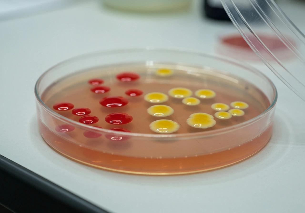
MSA, also called Chapman agar, has a straightforward three-part logic. The formulation contains 75 g/L sodium chloride (7.5% w/v), 10 g/L D-mannitol, 0.025 g/L phenol red indicator, and approximately 15 g/L agar at a final pH of around 7.4. Each component plays a specific role.
- 7.5% NaCl (selective agent): This concentration is far higher than most physiological media. It kills or inhibits the vast majority of bacteria, allowing only salt-tolerant (halotolerant) organisms to form colonies.
- D-mannitol + phenol red (differential system): Organisms that ferment mannitol produce acid, dropping the pH and turning the phenol red indicator from red-pink to yellow. Non-fermenters leave the medium unchanged or make it turn pink-red.
- Agar base: Provides the solid support. The medium is transparent enough to read color changes clearly around and under colonies.
Commercial products from BD BBL, Thermo Fisher Remel, and Neogen all use the same Chapman formulation and explicitly describe MSA as selective for staphylococci. In short, mannitol salt agar will only grow bacteria that are halotolerant (salt-tolerant), primarily staphylococci, and thus is not suitable for organisms like Proteus. The high salt concentration is the reason: staphylococci tolerate 7.5% NaCl comfortably, while most other bacteria, including the Enterobacterales family to which Proteus belongs, cannot. This is the core reason MSA is not used to screen for Proteus in any standard protocol.
Key lab traits of Proteus vulgaris that predict its behavior on selective media
Understanding why Proteus vulgaris behaves the way it does on MSA requires a quick look at its fundamental characteristics. These traits also explain why certain other media are far better choices for isolating it.
- Gram-negative rod: Proteus vulgaris is a Gram-negative member of the order Enterobacterales. This alone places it in a category that MSA is designed to exclude.
- Limited halotolerance: Most Proteus strains can grow at roughly 4% NaCl but are frequently inhibited at 6.5% and above. The 7.5% NaCl in MSA is well above the tolerance ceiling for the majority of clinical and food-environment isolates.
- Rapid urease production: P. vulgaris hydrolyzes urea quickly, making urease positivity one of its most useful identification markers in the lab.
- Phenylalanine deaminase (PAD) positive: This biochemical trait, along with H2S production on TSI agar, further distinguishes Proteus from other Gram-negative rods.
- Indole-positive: P. vulgaris is characteristically indole-positive, which distinguishes it from the closely related P. mirabilis (indole-negative). This is a key species-level discriminator.
- Motile and swarming: P. vulgaris produces flagella and swarms across moist agar surfaces in concentric rings, which can make colony isolation difficult on standard blood agar.
These traits collectively mean that Proteus vulgaris belongs to a completely different ecological and physiological category than staphylococci. It is not built for high-salt environments, and its defining characteristics, fast urease activity, swarming motility, and Gram-negative cell wall structure, have no relevance to growth on MSA.
What actually happens when you plate Proteus vulgaris on MSA
In a standard culture setup, Proteus vulgaris will show no growth or only trace growth on MSA after 24 to 48 hours of incubation at 35-37°C. The 7.5% NaCl creates osmotic stress that the organism cannot overcome well enough to form visible colonies. There is no color change in the medium because no acid is produced from mannitol fermentation (the organism is not fermenting mannitol even if a few cells survive).
That said, a few caveats exist. Research dating back to a 1964 Nature letter by P. G. D. Naylor showed that some P. vulgaris strains demonstrate increased growth tolerance with rising NaCl concentrations under certain broth conditions, and blank" rel="noopener noreferrer">environmental surveys have recovered halotolerant Proteus strains from saline habitats such as salt lakes and salterns. Naylor (1964) reported that Proteus vulgaris tolerated and in some broth conditions showed increased growth with rising NaCl concentrations (Effect of Sodium Chloride, Dulcitol and Glucose on the Growth of Proteus in Shaken and Static Broth Cultures, P. G. D. Naylor, Nature (1964)) blank" rel="noopener noreferrer">Effect of Sodium Chloride, Dulcitol and Glucose on the Growth of Proteus in Shaken and Static Broth Cultures — P. G. D. Naylor, Nature (1964). These are genuine exceptions rather than the expected outcome. In a diagnostic or food-safety context, you should not count on Proteus growing on MSA, and you should not rely on a negative MSA result to rule it out.
If you do see any growth on MSA and suspect Proteus, the morphology will not match the typical swarming pattern you would see on blood agar, and the medium will likely remain red (no mannitol fermentation). Confirmatory testing would be essential before drawing any conclusion.
How Proteus compares across four common media
To put MSA in context, here is how Proteus vulgaris typically behaves on the four media most relevant to its detection and identification.
| Medium | Expected Growth | Colony Appearance / Notes |
|---|---|---|
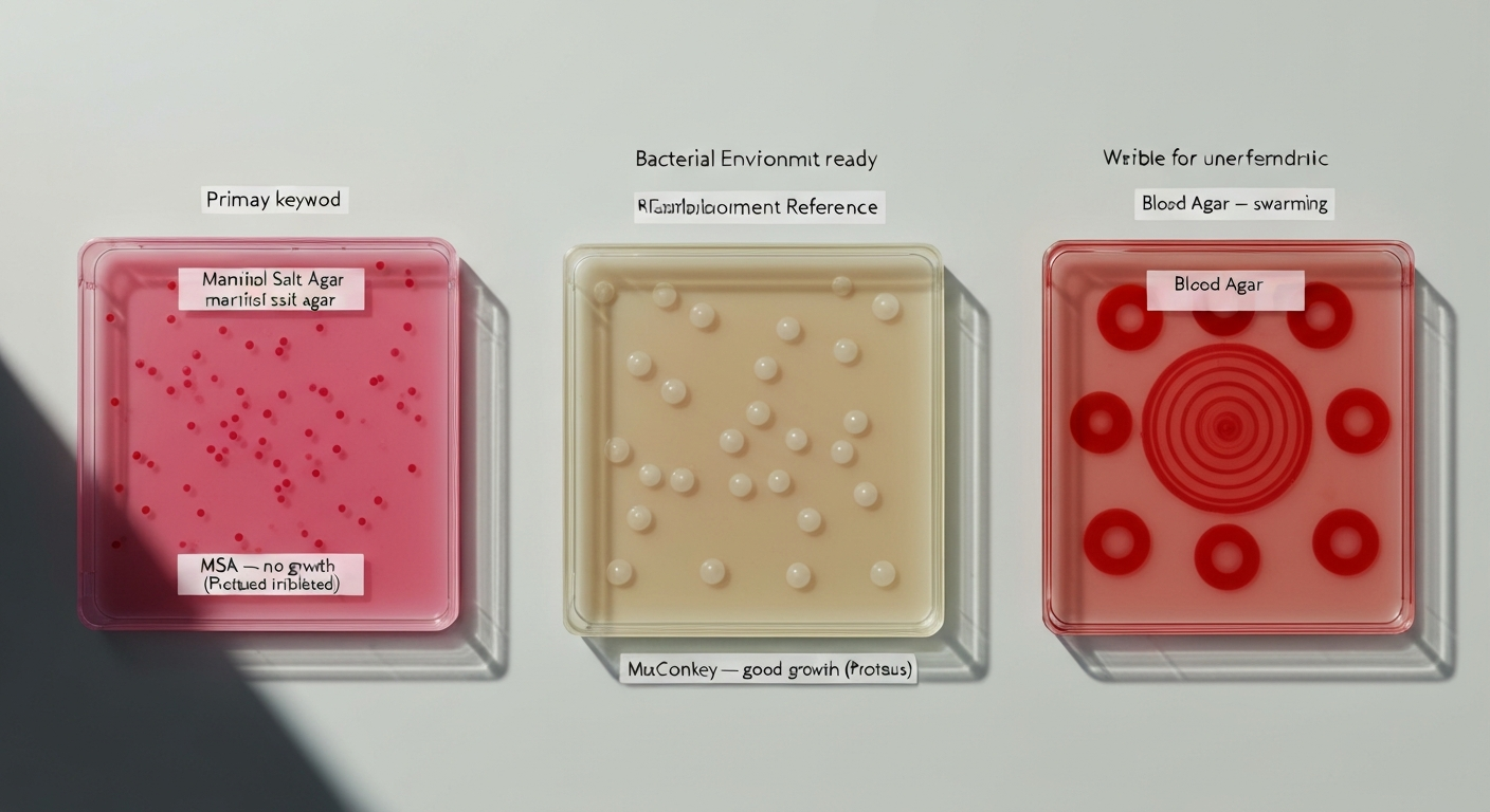
| Mannitol Salt Agar (MSA) | None or trace (inhibited) | No visible colonies in most cases; medium stays red; not a suitable isolation medium for Proteus |
| MacConkey Agar | Good growth | Pale/colorless (non-lactose fermenter); bile salts inhibit swarming, making individual colonies visible; standard selective medium for Proteus isolation |
| Blood Agar (BAP) | Good growth | Swarming colonies in concentric rings; may show hemolysis; swarming can overrun other colonies, complicating mixed-culture work |
| CLED Agar | Good growth | Pale yellow or colorless colonies; electrolyte-deficient formulation inhibits swarming; standard urine culture medium ideal for Proteus isolation from clinical specimens |
The contrast between MSA and the other three media is stark. Where MSA blocks Proteus with salt, MacConkey and CLED actively support its growth while suppressing swarming through their respective chemical compositions (bile salts on MacConkey; electrolyte deficiency on CLED). If you're also considering non-Enterobacterales like Pseudomonas, see the short note on whether Pseudomonas grows on MacConkey agar does Pseudomonas grow on MacConkey agar?. For specifics about Salmonella's behavior on this medium, see does salmonella grow on macconkey agar which discusses Salmonella growth and lactose fermentation on MacConkey. Blood agar supports the fullest expression of Proteus growth but makes swarming a practical problem. For anyone comparing Proteus behavior across selective agars, its expected performance on MacConkey is a useful point of reference alongside these MSA results. See 'does proteus vulgaris grow on macconkey agar' for specific expectations and troubleshooting.
A practical workflow for detecting and identifying Proteus from environmental or food samples
If you are trying to find Proteus vulgaris in an environmental or food sample, skip MSA entirely. Here is a straightforward workflow based on standard clinical and food-microbiology methods.
- Primary plating: Inoculate MacConkey agar and blood agar (or CLED if this is a urine or environmental water sample). Incubate at 35-37°C for 18-24 hours. MacConkey and CLED both inhibit swarming and allow individual colony selection.
- Colony selection: On MacConkey, pick pale or colorless non-lactose-fermenting colonies. On CLED, look for pale yellow or colorless colonies. On blood agar, watch for the characteristic swarming pattern with concentric rings.
- Rapid urease test: Streak or spot-inoculate a urea agar slope or Christensen's urea broth. Proteus vulgaris produces a fast, strong positive (color change within 1-4 hours), which is one of the most reliable quick flags for this genus.
- Phenylalanine deaminase (PAD) test: A positive PAD result (green color with ferric chloride) confirms membership in the Proteus-Providencia-Morganella group.
- Indole test: Add Kovac's reagent to a tryptone broth culture. Proteus vulgaris is typically indole-positive (pink-red ring), distinguishing it from P. mirabilis (indole-negative).
- TSI (Triple Sugar Iron) agar: Expect an alkaline slant/acid butt with H2S blackening, consistent with Proteus.
- Confirmatory ID: Use an API 20E strip, VITEK 2, MALDI-TOF, or equivalent automated system for definitive species-level identification, especially important when reporting to food safety authorities or in clinical settings.
Incubation at 35-37°C for 18-24 hours is appropriate for primary isolation. If swarming is a problem on blood agar, transfer suspicious zones to MacConkey or CLED for subculture before biochemical testing.
Controlling Proteus swarming during culture work
Proteus swarming is one of the most practically disruptive behaviors in routine bacteriology. On blood agar, the organism spreads in waves across the entire plate, potentially obscuring co-inhabitants in a mixed culture. There are several reliable ways to manage it.
- Use CLED agar: The electrolyte-deficient formula was specifically developed to inhibit Proteus swarming without sacrificing growth yield. It is the most consistently effective option for urine and environmental samples.
- Use MacConkey agar: Bile salts in MacConkey reliably suppress swarming, and the medium is widely available. Colonies are distinct and countable.
- Increase agar concentration: Raising the agar content to 5-6% instead of the standard 1.5% physically restricts swarming, though this approach is less practical in routine workflows.
- p-Nitrophenylglycerol (pNPG): Used in some research settings as a chemical inhibitor of swarming; not commonly required in routine clinical or food-safety labs.
- Subculture from swarming edge: If you are working with blood agar and swarming has occurred, you can still pick individual cells from the leading edge of a swarm ring for subculture onto a swarming-inhibitory medium.
In a food-safety or environmental monitoring context, the simplest rule is to never rely on blood agar alone when Proteus is a suspected organism in a mixed sample. Start with CLED or MacConkey and use blood agar only as a secondary confirmation plate.
What a negative MSA result actually tells you in food-safety sampling
In environmental or food-safety sampling, MSA is used to screen for halotolerant staphylococci. If you run an MSA plate and get no growth, it tells you that no significant population of staphylococci was present. It tells you absolutely nothing about Proteus vulgaris, because Proteus is expected to be negative on MSA whether it is present in the sample or not.
This is an important interpretation pitfall. A food-safety professional screening a surface swab on MSA and finding no growth should not conclude the surface is free of Gram-negative pathogens. Proteus, Salmonella, E. coli, and other Enterobacterales will all fail to grow on MSA under normal conditions. Separate plating on MacConkey or appropriate enrichment broths (such as Selenite F or Buffered Peptone Water) is required to screen for those organisms.
When Proteus contamination is specifically suspected, for example in a processed meat facility, a water system, or a hospital food-service environment, MacConkey agar or CLED should be the primary screening medium, not MSA. Enrichment steps can improve sensitivity for low-level contamination before plating.
Limitations and interpretation pitfalls to keep in mind
No selective medium is perfect, and MSA comes with a specific set of caveats that matter in real-world sampling.
- Rare halotolerant Proteus strains: Environmental isolates from high-salt habitats can occasionally grow at 7.5% NaCl. If you are sampling saline or brined food environments, do not treat a negative MSA as definitive evidence that Proteus is absent.
- False positives on MSA: Other halotolerant organisms, including some coagulase-negative staphylococci (CoNS) that ferment mannitol, can produce yellow halos on MSA and be mistaken for S. aureus. Coagulase testing or definitive molecular identification is always required to confirm S. aureus after MSA growth.
- False negatives on MSA: Some S. aureus variants are mannitol-use-deficient or slow-fermenting, so they may grow on MSA without producing the expected yellow color change. Relying solely on color change for identification is unreliable.
- Mixed cultures: In complex samples (food matrices, environmental swabs), a dense background of halotolerant bacteria can mask or inhibit the growth of target organisms, or vice versa.
- Timing: Reading MSA plates at 24 hours versus 48 hours can give different colony counts, especially for slow-growing organisms. Standardize your read time.
- Medium variability: Slight batch-to-batch variation in commercial MSA plates can affect selectivity. Always run positive and negative controls when using new lots.
Selective agar choices go beyond MSA and Proteus
The question of whether Proteus vulgaris grows on MSA is part of a broader practical topic: how do you choose the right selective medium for the organism you are actually looking for? Each selective agar works by exploiting a specific physiological characteristic of the target organism or the target group.
MSA exploits halotolerance to select for staphylococci. MacConkey agar exploits bile salt and crystal violet tolerance to select for Gram-negative Enterobacterales while inhibiting Gram-positive bacteria. This means that questions about whether Gram-positive bacteria can survive on MacConkey, or whether organisms like Pseudomonas or Burkholderia cepacia grow on MacConkey, follow the same underlying logic: each comes down to which specific selective pressures the organism can withstand. If you need specifics about whether Burkholderia cepacia grows on MacConkey agar, consult the guidance titled does burkholderia cepacia grow on macconkey agar.
For Proteus specifically, MacConkey is both a suitable growth medium and a swarming-control tool, which makes it especially useful. The key takeaway for any sampling program is to match your medium to your target organism's known tolerances before you plate, not after you get unexpected results.
FAQ
Does Proteus vulgaris grow on Mannitol Salt Agar (MSA)?
Generally no, or only very poor/strain‑dependent growth. MSA contains about 7.5% NaCl which selectively favors halotolerant staphylococci; most Proteus vulgaris isolates (a Gram‑negative Enterobacterales member) are inhibited by that high salt and will not produce typical colonies on MSA.
Why is MSA inhibitory to Proteus vulgaris?
MSA is formulated with a high sodium chloride concentration (≈7.5% w/v) as the selective agent and contains mannitol plus phenol red to differentiate mannitol fermenters. The high salt level inhibits many non‑staphylococcal bacteria, including most Proteus strains that lack strong halotolerance.
Are there exceptions — can any Proteus strains grow on MSA?
Yes — salt tolerance is strain‑dependent. Some environmental or atypical Proteus isolates reported in the literature tolerate elevated NaCl and in rare cases may grow on high‑salt media. However, these are exceptions; clinical and routine practice treats MSA as inhibitory to most Proteus isolates.
What are the typical laboratory traits of Proteus vulgaris relevant to identification?
Proteus vulgaris is a Gram‑negative, facultative anaerobe in Enterobacterales; it is motile (swarming on many non‑inhibitory agar plates), urease positive, phenylalanine deaminase (PAD) positive, usually indole positive, and can produce H2S on appropriate media. It is typically recovered on blood agar, MacConkey, or CLED rather than on MSA.
How do expected results for Proteus vulgaris compare across common media (MSA vs MacConkey vs blood agar vs CLED)?
MSA: usually no growth or very poor growth; if growth occurs, colonies are atypical and unreliable for ID. MacConkey: supports growth of Proteus (non‑lactose fermenters give colorless colonies) and contains bile salts that suppress swarming. Blood agar: supports robust growth but swarming can overrun plates. CLED: supports growth and is formulated to reduce Proteus swarming (electrolyte‑deficient), making it useful for urine cultures.
What culture and incubation conditions are recommended to isolate and identify Proteus vulgaris?
Incubate plates at 35–37°C for 18–24 hours (longer if needed). For isolation use blood agar, MacConkey, or CLED. To control swarming, use CLED or MacConkey, increase agar concentration, or plate from a single very dilute inoculum; if blood agar is used, subculture promptly to differential/biochemical media. After isolation, confirm with biochemical tests (urease, indole, PAD, TSI/H2S) or a validated rapid/automated ID method.
Next Article
Mannitol Salt Agar Will Only Grow Bacteria That Are Salt Tolerant
Explains that mannitol salt agar selects salt tolerant bacteria and differentiates mannitol fermenters via color change.