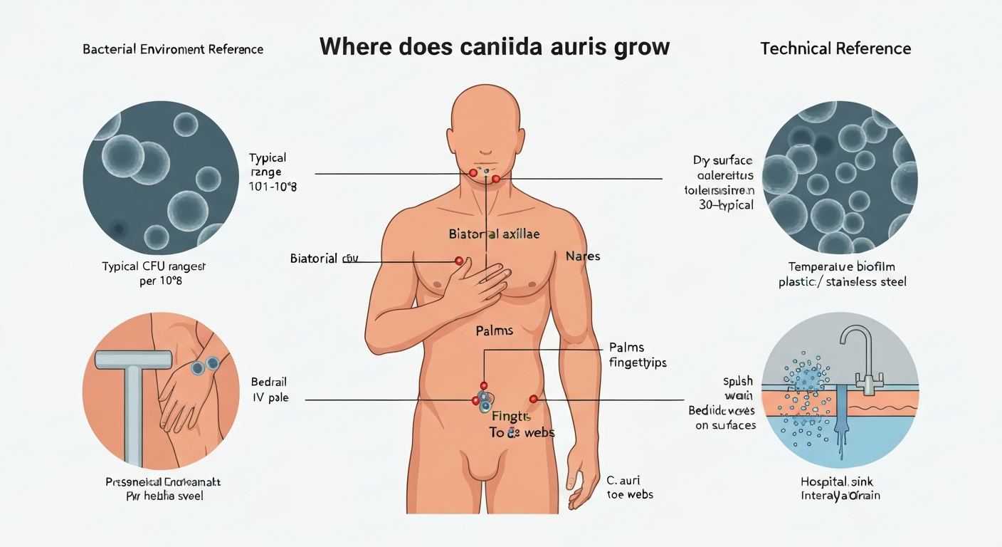
Candida auris grows primarily on human skin and in the healthcare environment. In colonized patients, it concentrates in the axilla, groin, nares, palms, fingertips, and toe webs. Outside the body, it persists on plastic, vinyl, stainless steel, and other hard surfaces in patient-care rooms for days to weeks, and it forms dry-surface biofilms that make it significantly harder to remove than most other Candida species. It is not a recognized foodborne pathogen, and there is no current evidence it grows in or on foods under normal handling conditions.
Where Does Candida auris Grow: Habitats, Surfaces, Risks

Marcus Reeves
30 Jul 2026
Scope, audience, and how to use this reference
This article is a technical reference for food-safety professionals, infection-prevention staff, clinical microbiologists, educators, and informed consumers who want a factual answer to where Candida auris grows and what environmental conditions support that growth. It covers human colonization sites, healthcare surface reservoirs, environmental growth requirements, laboratory culture conditions, biofilm behavior, and practical guidance for environmental control. It also addresses the question of food safety risk, which is low but worth understanding clearly.
Use this article as a starting point, not a clinical protocol. For active outbreak management or patient-care decisions, consult your institution's infection-prevention team, clinical microbiology laboratory, or public-health authority. This reference is organized so you can read straight through or jump to the section most relevant to your role.
Brief biology relevant to environmental survival
Candida auris is a multidrug-resistant yeast, first described in 2009, that belongs to the Candida haemulonii complex. Unlike most other Candida species, it is unusually thermotolerant, capable of growing at human body temperature and well above it. It reproduces primarily by budding as yeast cells rather than forming hyphae, and it secretes an extracellular matrix that allows it to build adherent biofilms on abiotic surfaces. It also expresses a species-specific adhesin called SCF1 that actively promotes attachment to skin and device materials.
These traits explain its healthcare dominance. Most environmental fungi are eliminated by body temperature, killed quickly on dry surfaces, or eradicated by standard antifungals. C. auris passes all three of those filters more reliably than most fungal pathogens, which is why it has become a global infection-control problem in hospitals and long-term care settings rather than a community or food-production concern.
Where C. auris colonizes the human body
Colonization means C. auris is present and detectable on a body site without necessarily causing infection or symptoms. The organism can colonize multiple sites simultaneously, and its distribution across the body has been mapped carefully in outbreak investigations.
Primary colonization sites
The CDC recommends bilateral axilla-groin composite swabs as the standard surveillance specimen in healthcare settings. That reflects epidemiological practicality, but it does not mean those are the only sites. A detailed quantitative study in a ventilator-capable skilled nursing facility found the anterior nares to be the most densely colonized body site, with a median peak abundance of approximately 10^8.2 CFU in positive patients, following a bimodal distribution. The axilla, inguinal folds, palms, fingertips, and toe webs were also consistently positive. Colonization burdens across all sites span a wide range, roughly 10^1 to 10^8 CFU per swab, so even low-burden sites matter for transmission.
Including nares swabs alongside the standard axilla/groin composite substantially increases detection sensitivity. For surveillance purposes, the tradeoff is cost and patient discomfort versus missing a colonized individual who is positive only in the nares. Risk-stratified programs at high-burden facilities often add nasal swabs precisely because of that high-density reservoir.
Duration of colonization
C. auris colonization can last months to years, and in some cohorts detection fluctuates over time without ever definitively clearing. The CDC notes that re-screening known colonized patients is generally not recommended because colonization may persist indefinitely. This persistence has direct implications for environmental contamination: a long-term care resident who remains colonized for two years continuously seeds their environment with viable cells, regardless of whether an active infection is present.
Wound sites, mucosa, and indwelling devices
C. auris can colonize wounds, central venous catheters, urinary catheters, and endotracheal tubes. Device-associated colonization carries clinical significance because biofilm formation on device surfaces creates a protected reservoir from which cells can seed the bloodstream. Mucosal sites, including the respiratory tract, have been documented as colonization reservoirs, which is consistent with the high nares burdens observed in quantitative studies.
Skin colonization dynamics: epidermis, dermis, and the resident vs transient flora question
Skin colonization by C. auris occurs in the epidermis, the outermost layers of the skin, not in the dermis. The dermis is a deeper, living tissue layer separated from the external environment by the epidermal barrier, and healthy intact dermis does not harbor surface colonizers. For details on whether resident flora can grow in the dermis, see the related entry "Does resident flora grow in the dermis.". This distinction matters practically: colonization screening swabs sample the skin surface and superficial layers, not deep tissue. Deep tissue invasion represents infection, not colonization, and is a different clinical scenario.
In microbiology, skin microorganisms are broadly divided into resident flora (organisms stably integrated into the skin microbiome over time) and transient flora (organisms present temporarily from environmental contact that can be removed by handwashing). C. auris occupies an unusual position. It behaves more like a persistent colonizer than a transient contaminant. Unlike transient flora, it is not reliably removed by standard hand hygiene, and colonized patients retain it across multiple body sites for extended periods. Yet it does not appear to be a true resident organism in the classic sense: it is not a normal component of healthy human skin flora and is not found in non-healthcare community populations at meaningful rates. It is essentially a healthcare-acquired persistent colonizer that mimics resident flora behavior once established. For a deeper look at where resident and transient flora are found across anatomical sites, those topics are covered in related reference entries on this site. For details on typical habitats and anatomical sites, see the related entry on where does transient flora grow.
Healthcare and built-environment reservoirs
Environmental contamination in rooms occupied by colonized or infected patients is well-documented. Outbreak investigations across the United States and internationally have cultured C. auris from bedrails, doorknobs, call buttons, portable equipment, thermometers, blood pressure cuffs, ventilators, IV poles, bedside tables, chairs, sinks, drains, and floors. The density of environmental contamination correlates with patient colonization burden and time in the room.
Sinks and drains are particularly important reservoirs. Drain biofilms are difficult to reach with standard terminal cleaning and can serve as a sustained source of recontamination after a room is cleaned. This means that even after a patient is discharged and the room is cleaned, a heavily contaminated drain can reseed surfaces brought into contact with drain water splash. Healthcare facilities dealing with persistent environmental contamination should treat sink drains as primary targets, not secondary ones.
Environmental growth requirements and tolerances
Understanding the conditions under which C. auris actually grows versus merely survives helps prioritize both prevention and remediation. Growth and persistence are different: an organism can be viable on a surface without actively multiplying. The thresholds below reflect current laboratory evidence, with notes where the data are limited or strain-dependent.
Temperature
C. auris grows robustly at 30 to 37°C, making it well-adapted to both human body temperature and typical healthcare room temperatures. Most isolates show substantial growth at 40 to 42°C, and some retain attenuated growth at temperatures approaching 47°C on plate assays. This upper tolerance is clade- and strain-dependent: not every isolate behaves identically, and colony size typically diminishes as temperature increases above 40°C. This thermotolerance exceeds most other Candida species and is considered one of the reasons C. auris succeeded as a human pathogen in warm-blooded hosts.
pH
Most C. auris isolates show no growth at pH 2 and are effectively inhibited by strongly acidic conditions. At the alkaline end, growth has been documented at pH values as high as 13 on plate assays, which is exceptionally high alkaline tolerance for a yeast. This is practically relevant: standard alkaline laundry detergents and some cleaning solutions may not eliminate C. auris from fabrics and textiles based on pH alone. Effective decontamination of linens requires validated laundering processes that combine temperature and appropriate chemistry, not just high pH.
Moisture and water activity
C. auris can persist on both dry and moist surfaces, which distinguishes it from many other fungi that require elevated water activity to survive for extended periods. Experimental work shows higher survival populations on wet metal surfaces, while separate studies demonstrate long survival on dried plastics. Persistence is surface- and context-dependent, meaning no single moisture threshold reliably predicts when the organism dies off. Many published persistence studies were conducted at approximately 25°C and moderate relative humidity (40 to 60%), which corresponds to typical hospital room conditions. Direct comparative RH-gradient experiments remain sparse in the literature, so specific minimum water-activity thresholds for growth versus survival are not yet well-defined for this organism.
Oxygen preference and nutrient requirements
C. auris is a facultative anaerobe, growing preferentially in aerobic conditions but capable of surviving in reduced-oxygen environments. For nutrient requirements, it grows on standard yeast-supporting substrates including glucose and other fermentable carbohydrates. There is no evidence it requires unusual substrates for environmental persistence; it can subsist on trace organic matter on surface films, which is part of why environmental contamination is sustained without visible organic soil.
| Parameter | Range / Tolerance | Notes |
|---|---|---|
| Temperature (growth) | 30–42°C typical; some isolates up to ~47°C (attenuated) | Strain and clade-dependent; thermotolerance exceeds most Candida spp. |
| Temperature (survival on surfaces) | Persists at room temperature (~20–25°C) | Laboratory studies conducted at ~25°C confirm multi-week persistence |
| pH (growth) | No growth at pH 2; growth documented up to pH 13 | High alkaline tolerance relevant to detergent/laundry exposures |
| Water activity / moisture | Persists on both dry and moist surfaces | Direct minimum water-activity data limited; surface-dependent |
| Relative humidity | Persists at ~40–60% RH under lab conditions | RH-gradient experimental data sparse; typical hospital RH tolerated |
| Oxygen preference | Facultative anaerobe; aerobic preferred | Survives in reduced-oxygen environments |
| Nutrient substrate | Standard yeast substrates; trace organics sufficient for persistence | No unusual nutrient requirements identified |
Materials, surfaces, and devices that support growth or long-term survival
Laboratory deposition studies have established that C. Survival, Persistence, and Isolation of the Emerging Multidrug‑Resistant Pathogenic Yeast Candida auris on a Plastic Health Care Surface (Journal of Clinical Microbiology, 2017) reported that C. auris remained culturable on plastic surfaces for at least 14 days under those conditions A controlled laboratory study reported that C. auris remained culturable on plastic healthcare surfaces for at least 14 days at ~25°C and ~57% relative humidity.. auris remains culturable on nonporous plastic healthcare surfaces for at least 14 days at 25°C and approximately 57% relative humidity. Metabolic viability, measured by esterase activity, persisted for up to 28 days, meaning viable-but-nonculturable cells remained even after standard culture methods stopped detecting them. This is important for environmental sampling: a negative culture result from a surface does not guarantee the organism is absent or nonviable.
Published experimental and review literature reports C. auris survival across plastic, stainless steel, vinyl, and glass for periods ranging from days to approximately three weeks or longer under healthcare-like conditions. Fabric and textile surfaces have received less experimental attention, but the organism's alkaline pH tolerance means standard alkaline detergent laundering cannot be assumed sufficient on its own.
Biofilm formation on abiotic surfaces
C. auris forms dry-surface biofilms (DSBs) on abiotic surfaces, a trait with major infection-control implications. These biofilms consist mainly of yeast cells embedded in extracellular matrix polysaccharides and confer substantially increased tolerance to cleaning agents, including sodium hypochlorite. A surface that appears clean visually may harbor a biofilm-protected population that is not eliminated by a single disinfection event.
On medical device materials including polystyrene, silicone, polyurethane, PVC, and catheter materials, C. auris forms adherent biofilms that show high tolerance to antifungals. The SCF1 adhesin promotes both initial surface attachment and subsequent biofilm development, and its expression appears to be upregulated in device-relevant conditions. This means that catheters, endotracheal tubes, and other indwelling devices are not just passive reservoirs but active biofilm scaffolds.
Can Candida auris grow in or on foods?
Based on current evidence, C. auris is not a foodborne pathogen and there is no documented evidence that it grows in or on foods under normal agricultural, processing, or handling conditions. It is not associated with food production environments, and no outbreaks or cases of C. auris infection have been linked to food consumption. This contrasts with mycotoxin-producing molds, which actively colonize grain, nut, fruit, and processed food substrates and represent a genuine, well-characterized food-safety risk. For more detail on the food substrates that mycotoxins can grow on, see the related mycotoxin reference entry.
The reason C. auris has not emerged as a food concern likely comes down to transmission ecology. It spreads person-to-person and via contaminated healthcare surfaces, not through the food supply. Its colonization niche is healthcare settings with vulnerable immunocompromised hosts, not the broader environment or food chain. That said, the formal evidence base is limited because systematic testing of food substrates for C. auris growth has not been a research priority. For food-safety professionals, the current risk assessment is low, but awareness is appropriate as the organism's global distribution expands.
Laboratory culture and detection conditions
In the laboratory, C. auris is typically cultured on Sabouraud dextrose agar (SDA), CHROMagar Candida Plus, or C. auris-selective supplemented agar at 35 to 37°C for 24 to 48 hours for primary growth, with up to 72 hours recommended for environmental samples where cell numbers may be low. Some protocols use RPMI 1640 or yeast nitrogen base media for susceptibility testing. For environmental swabs, enrichment broth steps before plating can improve recovery from low-burden surfaces.
Identification is a well-documented challenge. C. auris is misidentified by many commonly used automated identification systems, including older VITEK 2 databases and API 20C AUX strips, as Candida haemulonii, Candida famata, Saccharomyces cerevisiae, or other organisms. MALDI-TOF mass spectrometry with an updated reference database is currently the most reliable routine identification method. Molecular methods including ITS sequencing or species-specific PCR provide definitive identification. Any laboratory using older phenotypic or automated platforms should verify that their system has C. auris in its current reference library before reporting a negative result as reliable.
How C. auris compares to mycotoxin-producing molds and Bacillus anthracis
Placing C. auris in comparative context helps clarify what makes it distinctive rather than treating it as a generic fungal or microbial hazard.
| Feature | Candida auris | Mycotoxin-producing molds (e.g., Aspergillus, Fusarium) | Bacillus anthracis |
|---|---|---|---|
| Primary habitat | Human skin; healthcare surfaces | Soil, grain, fruits, indoor damp environments | Soil; spores extremely persistent in soil for decades |
| Key substrate | Skin, device materials, abiotic surfaces | High-carbohydrate food substrates, organic debris | Soil organic matter; animal carcasses |
| Growth form | Yeast (budding); biofilm | Filamentous hyphae; spores | Rod-shaped bacterium; endospore-forming |
| Foodborne risk | Not demonstrated | Well-established; mycotoxins contaminate grain, nuts, feeds | Low (rare gastrointestinal anthrax via contaminated meat) |
| Temperature range | 30–42°C typical; some strains to ~47°C | Species-dependent; many grow 20–35°C, some at lower temps | 10–45°C; sporulation favored in soil |
| Environmental persistence mechanism | Dry-surface biofilm; desiccation tolerance | Sporulation; mycotoxins chemically stable | Highly resistant endospores (decades in soil) |
| Primary public-health concern | Healthcare-associated invasive infections in immunocompromised patients | Mycotoxin ingestion (hepatotoxic, carcinogenic, immunosuppressive) | Inhalational and cutaneous anthrax; bioterrorism concern |
Mycotoxin-producing molds and C. auris both involve fungal biology, but they occupy almost entirely different niches and pose different hazards. Mold-associated mycotoxins are a food-safety and agricultural concern primarily through ingestion. C. auris is a healthcare infection-control concern through direct contact and device colonization. Bacillus anthracis, covered separately in this reference for anthrax growth conditions, represents a different kingdom entirely and is notable for spore-based environmental persistence that vastly exceeds even C. auris in duration and severity of environmental threat.
Practical environmental sampling: where to look and what to expect
For infection-prevention purposes, environmental sampling in a suspected C. auris-affected area should prioritize high-touch surfaces (bedrails, call buttons, overbed tables, doorknobs, IV poles), sink basins and drain surrounds, and surfaces of portable equipment that travels between patients. Floor samples near the bed are also worthwhile as secondary targets.
Sampling method matters. Nylon-flocked swabs moistened with neutralizing transport buffer recover more cells from dry surfaces than dry swabs alone. RODAC contact plates work well for flat nonporous surfaces. For drains, direct swabbing of the drain interior surface after partial disassembly yields better results than swabbing the drain opening alone.
Timing affects results. Sampling immediately after terminal cleaning underestimates baseline contamination; sampling before cleaning or 24 to 48 hours after occupancy begins gives a more representative picture. A negative culture result does not rule out the presence of viable-but-nonculturable cells, particularly on surfaces where biofilm may have formed. Interpret negative environmental samples conservatively when there is epidemiological reason to suspect C. auris is present.
Cleaning, disinfection, and prevention
Standard quaternary ammonium compound (quat) disinfectants show reduced efficacy against C. auris compared to most other Candida species, and they are not recommended as the primary product for C. auris environmental control. The CDC and most infection-control guidance recommend sporicidal agents or EPA-registered disinfectants specifically tested against C. auris. Sodium hypochlorite (bleach) at concentrations of 1,000 to 5,000 ppm with adequate contact time (typically 5 to 10 minutes) is effective on hard, nonporous surfaces.
Contact time is the most commonly overlooked factor in practice. A disinfectant applied and immediately wiped off does not have time to work. In clinical environments, staff should verify that surfaces remain visibly wet for the full labeled contact time before drying or reuse. This is especially important for high-touch surfaces and anything that was in direct contact with a colonized patient.
Sinks, drains, and drain surrounds require dedicated attention beyond standard room cleaning. Options include pouring a hypochlorite solution directly into drains, using drain-specific disinfectant products, or in persistent outbreak scenarios, physically removing and replacing drain components. Fabrics and soft surfaces present a greater challenge: laundering should use hot water at temperatures sufficient to kill the organism (at least 60 to 71°C) combined with an appropriate detergent, given that alkaline pH alone is insufficient based on the organism's pH tolerance profile.
Summary of disinfection recommendations
- Use EPA-registered disinfectants with demonstrated efficacy against C. auris or against Clostridioides difficile spores (which typically indicates sporicidal activity sufficient for C. auris).
- Avoid relying solely on quaternary ammonium compounds as the primary agent for C. auris rooms.
- Apply sodium hypochlorite at 1,000 to 5,000 ppm with a contact time of at least 5 to 10 minutes on hard nonporous surfaces.
- Address sinks and drains explicitly in terminal cleaning protocols, not just room surfaces.
- Ensure laundering of fabrics reaches at least 60 to 71°C; do not rely on alkaline detergent pH alone.
- Document and audit contact times during cleaning; this is the most frequent gap in disinfection practice.
- Conduct post-cleaning environmental sampling after high-risk room turnovers to verify efficacy.
When to consult clinical microbiology, infection prevention, or public health
Consult your clinical microbiology laboratory immediately if C. auris is identified or suspected in any patient specimen, because many laboratories need to activate enhanced identification protocols or confirm results with reference methods. Early communication prevents misidentification from propagating through the medical record.
Engage infection prevention when a single confirmed case is identified in a healthcare facility. C. auris is a reportable condition in many U.S. states, and public-health notification may be legally required. Your facility's infection-prevention team will initiate contact precautions, colonization screening of unit contacts, and enhanced environmental cleaning protocols.
When contacting any of these experts, bring the following information: the patient's clinical and exposure history, the body site from which C. auris was identified, the identification method used, the patient's unit and room assignment history, names of shared equipment or procedures, and the dates of symptom onset or specimen collection. This information substantially accelerates the investigation.
Key uncertainties and research gaps
Several important questions remain incompletely answered in the published literature, and recognizing them matters for risk-based decision making.
- Minimum water activity for growth versus survival on surfaces has not been precisely defined experimentally. Most published persistence studies were conducted at a single RH level rather than across a gradient.
- RH-gradient experiments specific to C. auris are sparse. Current guidance on moisture control is extrapolated from related yeasts and from the organism's demonstrated behavior at typical hospital RH ranges.
- Clade- and strain-level variation in thermotolerance, pH tolerance, and surface persistence is documented but not systematically mapped. Guidance developed from one clade may not fully apply to another.
- The foodborne risk assessment is currently based on absence of reported cases and ecological reasoning rather than systematic experimental inoculation studies of food substrates.
- Viable-but-nonculturable (VBNC) cells complicate environmental sampling interpretation. The prevalence, duration, and infectivity of VBNC C. auris on surfaces are not well characterized.
- Efficacy data for many EPA-registered disinfectants were generated against specific reference strains. Whether those results generalize across the diversity of clinical and environmental isolates in circulation is not fully established.
- The role of the community environment (non-healthcare settings, soil, water) in C. auris ecology and transmission is poorly understood despite its designation as an emerging pathogen.
For practical risk management, these gaps argue for using conservative assumptions: treat surfaces in known C. auris rooms as contaminated until confirmed otherwise, use sporicidal or validated fungicidal agents rather than relying on general disinfectants, and maintain colonization surveillance in high-risk populations even after apparent clearance. As the evidence base grows, guidance will continue to evolve, so checking for updates from the CDC Mycotic Diseases Branch and your regional public-health authority is worthwhile on a regular basis.
FAQ
Where does Candida auris typically colonize on the human body?
Candida auris is primarily a skin colonizer. Common screening and colonization sites are the bilateral axilla and groin (CDC recommends an axilla–groin composite swab for surveillance). Other frequently colonized sites include the anterior nares, palms/fingertips, toe webs, and perianal/inguinal regions. Quantitative burden varies widely by site and patient (reported ranges from ~10^1 to ~10^8 CFU per swab in outbreak studies). Colonization can be persistent (months to years) with fluctuating detection over time.
Does C. auris colonize deeper skin layers (dermis) or is it limited to epidermal/skin surface colonization?
Evidence indicates C. auris mainly colonizes the skin surface and superficial epidermal sites rather than the dermis. It behaves like other skin colonizers (resident or persistent flora in some people), although individual burden and duration vary. Distinguish resident (long‑term, stable colonization) versus transient flora (short‑lived contamination): C. auris can act as a persistent/resident colonizer in healthcare settings, which increases risk of environmental contamination and transmission.
Which healthcare and environmental surfaces support C. auris survival and growth?
C. auris survives and can persist (days to weeks) on many healthcare materials, including plastics (polystyrene, polyvinyl chloride), stainless steel, vinyl, glass, bedrails, doorknobs, thermometers, medical devices, and patient‑care equipment. Survival duration depends on surface type, moisture, temperature, and biofilm formation; experimental studies document culturability for ≥14 days on plastic and metabolic viability for longer under healthcare‑like conditions.
Can C. auris form biofilms and does that affect persistence?
Yes. C. auris forms adherent biofilms on abiotic surfaces and medical‑device materials (e.g., silicone, polyurethane, catheter materials). It also forms dry surface biofilms (DSBs). Biofilm growth increases environmental persistence and tolerance to antifungals and some disinfectants, making cleaning and disinfection less effective if biofilms are present.
What are the environmental growth requirements and tolerances of C. auris (temperature, pH, moisture, oxygen, nutrients)?
Temperature: most isolates grow at 30–37°C; many tolerate 40–42°C and some show attenuated growth up to ~47°C, with strain/clade variation. pH: sensitive to very acidic conditions (little/no growth at pH ~2) but relatively tolerant of alkaline conditions (growth observed at high pH values in lab tests). Moisture: can persist on both wet and dry surfaces; moist conditions may favor higher survival in some contexts, but long survival on dry plastics is well documented. Relative humidity: persistence documented in typical hospital RH (~40–60%); RH‑gradient data are limited. Oxygen: C. auris is a facultative aerobe/yeast that grows under standard laboratory aerobic conditions; it does not require specialized anaerobic conditions. Nutrients/substrates: grows on typical yeast media and can survive on low‑nutrient surfaces; biofilm formation and environmental survival do not require rich substrates but benefit from organic soil/bioburden.
Does C. auris grow in or on foods — is foodborne transmission a concern?
Available evidence does not support foods as a recognized reservoir or vehicle for C. auris transmission. C. auris is adapted to skin and healthcare environments; documented transmission is healthcare‑associated. Routine foodborne risk is currently considered negligible based on outbreak and environmental investigations. However, contamination of surfaces and equipment that contact food could theoretically transfer organisms if hygiene is poor — standard food‑safety hygiene measures mitigate that risk.
Next Article
Where Does Transient Flora Grow: Key Habitats and Controls
Where does transient flora grow: habitats, growth conditions and control measures for food safety and infection prevent