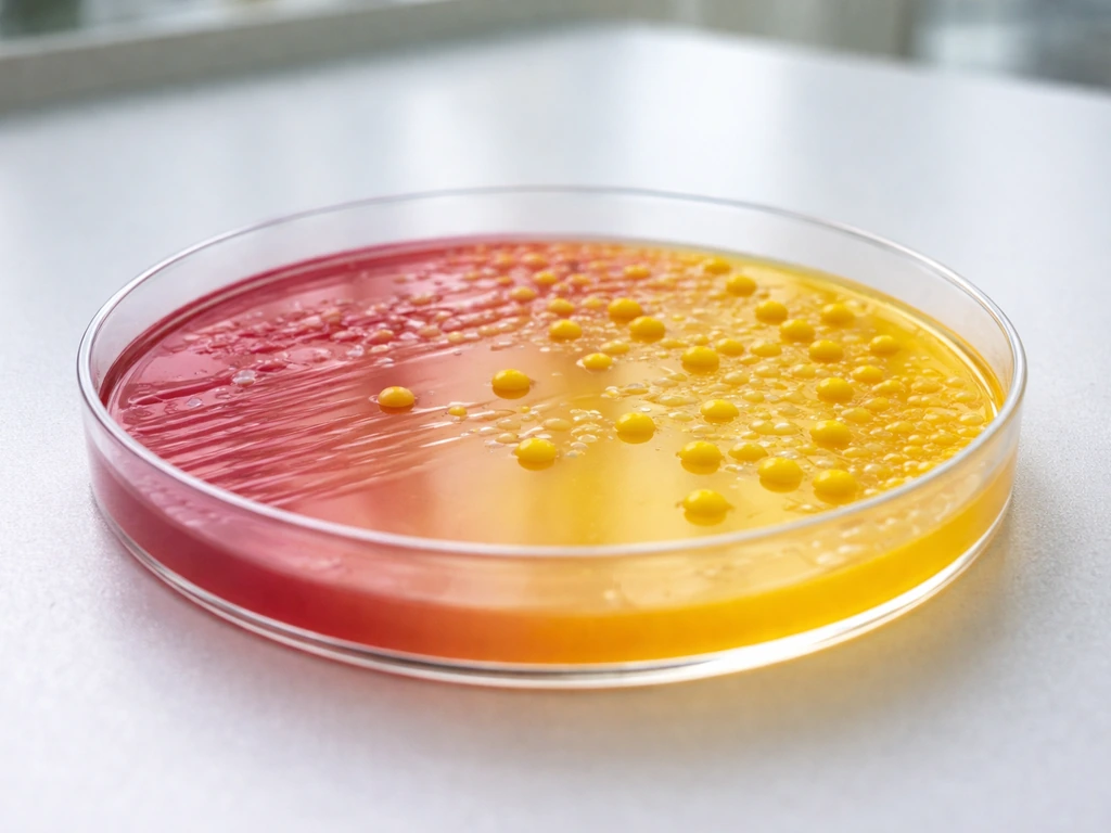
Yes, Micrococcus luteus will grow on Mannitol Salt Agar (MSA). The 7.5% NaCl in MSA is the medium's selective barrier, and M. luteus is halotolerant enough that most strains handle that salt concentration without much trouble. What it will not do is ferment mannitol, so the surrounding agar stays its original red-orange color. That distinction matters enormously in the lab: you can get a plate full of bright yellow M. luteus colonies and still have a completely negative mannitol result, which is a common source of misreading if you're not watching for it.
Does Micrococcus luteus Grow on MSA? Appearance & Tests

Marcus Reeves
13 Jul 2026
What MSA is actually made of and why it works as a selective medium
Standard MSA formulations (Oxoid, BD BBL, HiMedia) share a consistent recipe: 75 g/L sodium chloride (7.5% w/v), 10 g/L mannitol (1% w/v), and phenol red as a pH indicator. The agar is prepared to a starting pH of roughly 7.4 to 7.5 at 25°C. Those three components do different jobs.
The sodium chloride is the selective agent. At 7.5%, it creates an osmotic environment that most Gram-negative rods, streptococci, and other non-halotolerant organisms cannot survive. That leaves the plate largely open to halotolerant Gram-positive cocci, primarily staphylococci, which is exactly what the medium is designed to isolate. The mannitol and phenol red together form the differential system. If an organism acidifies the medium by fermenting mannitol, phenol red shifts from its original red-orange to yellow, coloring the agar around the colony. No fermentation means no acid, and the agar color stays unchanged.
The combination is efficient but not perfectly selective. Any organism tolerant of 7.5% NaCl will grow on MSA regardless of whether it ferments mannitol, and that is precisely where M. luteus enters the picture. The medium was designed with staphylococci in mind, but it does not chemically exclude other halotolerant members of the Micrococcaceae family.
Micrococcus luteus: the organism and the traits that matter here
M. luteus is a strictly aerobic, Gram-positive coccus classified within the Actinobacteria. You find it almost everywhere: human skin, soil, air, water, and a wide range of food-production environments. It forms tetrads or irregular clusters under the microscope, which is a useful first clue to its identity when working through Gram-positive coccus colonies on a plate.
The organism is mesophilic, with an optimal growth temperature around 28 to 30°C on standard media like tryptic soy agar or nutrient agar. Some strains tolerate temperatures up to around 45°C. For MSA work, the more important trait is its salt tolerance. Culture collection entries (ATCC 147, DSMZ/BacDive) and genomic studies of the Fleming reference strain document that many M. luteus strains survive and grow in media containing up to 10% NaCl. Since MSA uses 7.5%, the organism sits comfortably within that tolerance range for most strains, though there is genuine strain-to-strain variability.
Environmental isolates, particularly those recovered from saline or hypersaline settings, tend to sit at the higher end of salt tolerance. Clinical or skin-surface isolates may be slightly less robust at high salt, but in practice, growth on MSA is the expected outcome, not the exception.
Why M. luteus grows on MSA: the mechanism explained
MSA does not select against M. luteus because the organism has the same fundamental trait the medium is designed to select for: halotolerance. The 7.5% NaCl threshold is calibrated to exclude organisms with no salt tolerance while retaining staphylococci. M. luteus survives the same osmotic stress, so it passes through the selective filter.
Growth rate and colony size on MSA will typically be slower and smaller than what you see on nutrient agar or tryptic soy agar. The elevated salt imposes some osmotic burden even on tolerant strains. Published observations and lab experience consistently describe M. luteus on MSA as growing somewhat more slowly than Staphylococcus aureus under the same conditions, often producing smaller, drier colonies by the 48-hour read point. That said, growth is reliably visible, and at 72 hours colonies are usually easy to spot.
One nuance worth noting: the genome paper for the M. luteus Fleming strain reports that pigment synthesis (the carotenoid pathway responsible for the organism's characteristic yellow color) is diminished or abolished when NaCl concentrations exceed roughly 5%. The Fleming‑strain genome paper reports that pigment synthesis is diminished above ~5% NaCl (Genome sequence of Micrococcus luteus, osmotolerance and pigment note) Genome sequence of Micrococcus luteus — osmotolerance and pigment note. This means that on MSA, where NaCl sits at 7.5%, you may see paler, more cream-colored or off-white colonies rather than the bright yellow you would expect on standard nutrient agar. This detail matters for colony recognition, which is covered more fully below.
Mannitol fermentation: what M. luteus does (and does not) do
M. luteus does not ferment mannitol. It is a strictly aerobic organism and lacks the fermentative pathways needed to convert mannitol into acid end products under standard growth conditions. This is consistent across reference strains and environmental isolates described in the literature. On MSA, that means the phenol red indicator in the surrounding agar will not shift to yellow due to acid production. The agar around M. luteus colonies stays red-orange.
That non-fermentation result is actually the correct differential reading, but it can easily be misinterpreted if you are not careful about what you are looking at. The problem is that the organism itself produces yellow pigment under normal growth conditions, and even a muted version of that pigment on MSA can look, at a glance, like the agar-yellowing you would see from mannitol fermentation. The colony color and the medium color are two different things, and conflating them is one of the most reliable ways to get a false-positive mannitol reading.
What colonies look like on MSA
On standard nutrient agar at optimal temperature, M. luteus produces convex, circular, glistening, entire-edged colonies with a bright lemon-yellow color from carotenoid pigments. Some strains show a deeper orange-yellow; occasionally you see near-white variants, but yellow is the characteristic presentation most lab workers learn to associate with the organism.
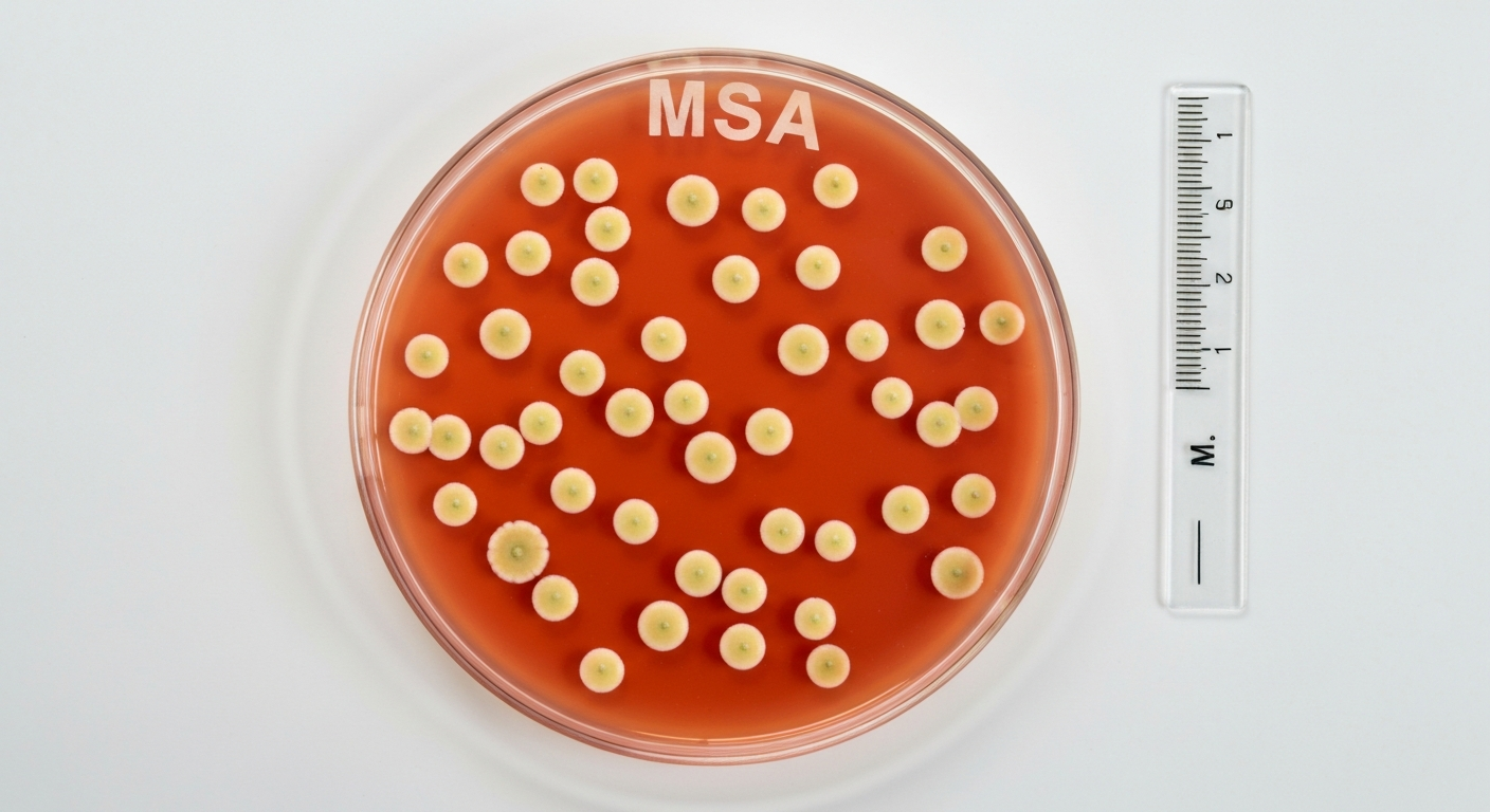
On MSA, expect those colonies to look different. Because high salt suppresses carotenoid synthesis, colonies are often pale yellow, cream, or off-white rather than the vivid yellow you see on nutrient agar. They tend to be smaller (1 to 2 mm at 48 hours is typical, compared to 2 to 3 mm on TSA at the same timepoint) and may appear slightly drier or more matte than S. aureus colonies on the same plate. The surrounding agar should remain red-orange, which is the critical visual cue.
Growth rate is noticeably slower than S. aureus under clinical incubation conditions (35 to 37°C), and even slower if an environmental isolate is being tested at those temperatures rather than the 28 to 30°C optimal range. If you are reading at 24 hours, you may see only pinpoint colonies or nothing at all for some strains. A 48-hour read is the minimum, and 72 hours gives you a more reliable picture.
The pigment problem: how yellow colonies mislead mannitol readings
This is probably the most practically important section of the article for anyone reading MSA plates in a real lab setting. The differential function of MSA depends on reading the color of the agar medium, not the color of the colony itself. Phenol red turns yellow when the pH drops below roughly 6.8 due to acid production from mannitol fermentation. That pH shift colors the agar around and under the colony, creating a halo effect that is the positive read.
M. luteus colonies, even when pale on MSA, still carry some yellow or cream pigment. A yellow colony sitting on red-orange agar can look, especially in lower light or to an inexperienced reader, like the agar itself has yellowed. Multiple lab manuals and teaching sources call this out explicitly as a common error. The rule is straightforward: look at the medium, not the colony. If the agar surrounding the colony remains red-orange, the mannitol result is negative regardless of how yellow the colony looks.
It is also worth noting that even if an isolate is genuinely yellow-pigmented on MSA (some strains retain partial pigment despite the elevated salt), the lack of an acid halo in the surrounding medium remains the definitive read. Do not let colony pigment substitute for a careful observation of agar color change.
Incubation conditions and when to adjust them
Commercial MSA specifications from Thermo/Oxoid, BD BBL, and HiMedia recommend incubating at 35 to 37°C for staphylococcal isolation, with a primary read at 48 hours. That protocol is designed for clinical isolates and S. aureus identification workflows, and it works well for those purposes.
For environmental or food-safety testing where M. luteus (or other environmental micrococci) may be present, consider extending incubation to 72 hours before calling negative results. If you know or suspect environmental isolates are involved, running a parallel plate incubated at 28 to 30°C gives you a better recovery rate and more representative colony morphology. The organism's optimal growth range sits closer to 30°C than 37°C, and forcing it to grow at the higher clinical temperature will slow it further and may reduce colony size to the point where it gets missed.
MSA is an aerobic medium, and this matters particularly for M. luteus, which is an obligate aerobe. Standard bench incubation with normal atmospheric oxygen is appropriate. Do not incubate MSA plates in anaerobic or CO2-enriched conditions for Micrococcus recovery; CO2 incubation is sometimes used for certain fastidious organisms but is not appropriate here and may suppress M. luteus growth.
| Parameter | Clinical/Standard Protocol | Environmental/Food Testing Adjustment |
|---|---|---|
| Temperature | 35–37°C | 28–30°C (or run parallel plates) |
| Primary read | 48 hours | 48 hours (extend to 72 hours before calling negative) |
| Atmosphere | Aerobic (ambient air) | Aerobic (ambient air); never anaerobic |
| Expected M. luteus colonies | Pale yellow/cream, small (may be underrepresented) | Brighter pigment, slightly larger, more typical morphology |
Confirming the isolate: a stepwise approach
Getting a colony on MSA is just the start. Because M. luteus grows on the medium but behaves very differently from S. aureus, confirmation testing is essential before drawing any conclusion about what you have. The following sequence covers the standard phenotypic battery and the more definitive molecular options.
Step 1: Gram stain and cell morphology
A Gram stain is the first move. Both M. luteus and staphylococci are Gram-positive cocci, so the stain alone will not separate them. But arrangement matters. M. luteus characteristically forms tetrads (groups of four) or irregular clusters of four to eight cells. S. aureus tends to form grape-like clusters. Tetrad arrangement is a useful early indicator, though it is not definitive on its own.
Step 2: Catalase test
Both M. luteus and staphylococci are catalase positive. A positive catalase result tells you that you have a member of one of these groups (as opposed to streptococci, which are catalase negative), but it does not differentiate between them. This step confirms you are in the right ballpark.
Step 3: Modified oxidase (microdase/TMPD) test
This is where the critical split happens. The microdase disk (TMPD, tetramethyl-p-phenylenediamine) tests for oxidase activity. Micrococcus species, including M. luteus, are modified oxidase positive, producing a blue-purple color on the disk within about 2 minutes. Most Staphylococcus species, including S. aureus, are modified oxidase negative. This single test correctly separates the genera in the vast majority of cases and should be considered standard for any catalase-positive Gram-positive coccus from MSA.
Step 4: Bacitracin (Taxo A) disk susceptibility
Low-dose bacitracin disks (0.04 units, the Taxo A format) produce a clear zone of inhibition around Micrococcus isolates. Staphylococci, including coagulase-negative species, are typically resistant and show no zone. This test is rapid, inexpensive, and provides reliable genus-level separation when used alongside the microdase result. The combination of microdase positive plus bacitracin susceptible makes an extremely strong phenotypic case for Micrococcus.
Step 5: Coagulase test (for S. aureus exclusion)
If your primary concern is ruling out S. aureus (the target organism for most MSA workflows), a coagulase test is straightforward. S. aureus is coagulase positive; M. luteus is coagulase negative, as are coagulase-negative staphylococci. A negative coagulase result does not confirm M. luteus on its own, but it eliminates S. aureus from the differential. Coagulase combined with the oxidase and bacitracin results gives you a solid working identification.
Step 6: Lysostaphin susceptibility
Lysostaphin cleaves the pentaglycine cross-bridges in staphylococcal peptidoglycan, which Micrococcus cell walls lack. Essentially all Staphylococcus strains lyse when exposed to lysostaphin, while Micrococcus strains are resistant. See Rapid lysostaphin test to differentiate Staphylococcus and Micrococcus species (PMC) for clinical validation that Staphylococcus strains are lysostaphin‑susceptible while Micrococcus strains are resistant. This test has been validated with high sensitivity and specificity across clinical strain panels and is particularly useful when you need to resolve a case where the microdase or bacitracin results are ambiguous.
Step 7: Molecular and MALDI-TOF confirmation
For food-safety investigations, outbreak contexts, or any situation where species-level identification needs to be definitive, 16S rRNA gene sequencing or MALDI-TOF mass spectrometry are the current gold standards. MALDI-TOF in particular gives rapid, genus- and species-level identification from a colony within minutes, and its reference databases now include M. luteus and related micrococci reliably. If your lab has access to MALDI-TOF, it is the most efficient endpoint when phenotypic results are equivocal.
Quick comparison: M. luteus vs S. aureus on MSA key tests
| Test | Micrococcus luteus | Staphylococcus aureus |
|---|---|---|
| Growth on MSA (7.5% NaCl) | Yes (most strains) | Yes |
| Mannitol fermentation | Negative (agar stays red-orange) | Positive (agar turns yellow) |
| Colony color on MSA | Pale yellow to cream or off-white | White to cream (gold on other media) |
| Gram stain arrangement | Tetrads / irregular clusters of 4 | Grape-like clusters |
| Catalase | Positive | Positive |
| Modified oxidase (microdase) | Positive | Negative |
| Bacitracin (Taxo A) susceptibility | Susceptible (zone present) | Resistant (no zone) |
| Coagulase | Negative | Positive |
| Lysostaphin lysis | Resistant | Susceptible |
What this means for food and environmental testing
In food safety work, MSA is typically deployed to detect and enumerate S. aureus. M. luteus is a common environmental contaminant on surfaces, air, and human skin, so it shows up on environmental swab plates regularly. Its presence on MSA is not inherently a food safety concern: M. luteus is not considered a recognized foodborne pathogen, and its growth on MSA is a selectivity limitation rather than a contamination alarm.
The practical risk is misidentification. An investigator who reads yellow M. luteus colonies on MSA as mannitol-positive may incorrectly flag the plate as S. aureus growth, triggering unnecessary responses. Running the confirmatory tests described above (at minimum, microdase and coagulase) before reporting any MSA result is good practice, not an optional extra.
For microbiologists working with the sibling topics in this reference set, similar selectivity questions arise with other organisms on differential media. For more on that topic, see does Bacillus subtilis grow on MSA for details about its salt tolerance and growth characteristics. Bacillus subtilis on MSA presents comparable salt tolerance considerations, while work with EMB agar for organisms like Alcaligenes faecalis or Serratia marcescens involves different selective chemistries but the same principle: a medium's selectivity has defined limits, and confirmation testing is always necessary. For more on how Alcaligenes faecalis behaves on eosin methylene blue (EMB) agar, see does Alcaligenes faecalis grow on EMB.
Alternatives to MSA and follow-up options
If your goal is specifically to isolate S. aureus with fewer interfering organisms, Baird-Parker Agar (BPA) is the more selective alternative used in food microbiology. BPA uses tellurite and egg yolk emulsion to create a system where S. aureus produces characteristic black colonies with clearing halos, and most micrococci do not interfere in the same visual way. It is the reference medium in many national and international food safety standards (including FDA BAM and ISO methods) for S. aureus enumeration from food.
Chromogenic S. aureus media (CHROMagar Staph aureus, MRSA-specific chromogenic plates) are another option for clinical and food labs needing high specificity. These formulations target S. aureus enzymatic activity and reduce non-specific growth more effectively than MSA alone.
If you specifically need to identify M. luteus rather than exclude it, standard tryptic soy agar or nutrient agar at 28 to 30°C is the right primary isolation medium. Colony pigment is more vivid, growth rate is optimal, and you are not working against the organism's salt tolerance when reading results.
Bottom line for lab practice
M. For the complementary question, does S. aureus grow on MSA, see the related discussion on S. aureus growth characteristics on Mannitol Salt Agar. luteus grows on MSA, does not ferment mannitol, and can produce yellow or pale yellow colonies that look similar to mannitol-positive agar yellowing at first glance. The way to avoid misidentification is to read agar color change (not colony color) for the mannitol result, and to run at minimum a modified oxidase test and coagulase test before reporting any MSA isolate. For food or environmental testing, extending incubation to 72 hours and considering a parallel plate at 30°C gives you the most complete picture. When the stakes are high, MALDI-TOF or 16S sequencing removes ambiguity entirely.
FAQ
Will Micrococcus luteus grow on Mannitol Salt Agar (MSA)?
Yes — many M. luteus strains are halotolerant and can grow on standard MSA (≈7.5% NaCl). Growth is strain dependent, but 7.5% NaCl is within reported tolerances for numerous strains.
Does Micrococcus luteus ferment mannitol and turn the MSA indicator yellow?
Generally no — M. luteus does not ferment mannitol. It can produce yellow pigmented colonies that may be mistaken for yellow medium acidification, but true mannitol fermentation is indicated by phenol‑red agar changing from red to yellow around colonies, not colony pigment alone. Pigment production itself is often reduced at higher NaCl concentrations.
What causes M. luteus to grow on MSA and why might it be misleading in differential reading?
MSA selects for halotolerant Gram‑positive cocci by using ~7.5% NaCl; M. luteus is halotolerant and strictly aerobic, so it can grow. The misleading element is colony pigmentation (bright yellow) which can be mistaken for agar acidification caused by mannitol fermentation. Because pigment may sit on top of unchanged red agar, visual colony color without checking surrounding agar is unreliable.
What will M. luteus colonies typically look like on MSA (appearance, pigment, margin)?
Expect small to medium, convex, glistening colonies often with yellow to golden carotenoid pigmentation. Pigment intensity can vary with strain and may be reduced at higher salt. Crucially, colonies often do not produce a yellow halo in the agar (no mannitol fermentation), so the medium surrounding colonies usually remains red.
What incubation conditions (temperature, time, atmosphere) are recommended when evaluating M. luteus on MSA?
Incubate aerobically. Use 24–48 hours as a routine window; inspect at 24 h and again at 48 h. Temperature: many staphylococcal protocols use 35–37°C, but for environmental/food samples incubating at 30°C (or parallel 30°C and 35–37°C) improves recovery of mesophilic Micrococcus strains and reduces misinterpretation due to temperature effects on pigment and growth rate.
What stepwise laboratory workflow and tests should be used to distinguish M. luteus from mannitol‑fermenting staphylococci?
Stepwise approach: 1) Observe MSA: note agar color change (phenol red) around colony, not colony pigment. 2) Gram stain: M. luteus = Gram‑positive cocci often in tetrads; Staphylococcus = Gram‑positive cocci in clusters. 3) Catalase: both are catalase positive. 4) Microdase/oxidase (TMPD) or modified oxidase: Micrococcus = positive; Staphylococcus = negative. 5) Bacitracin (Taxo A) disk: Micrococcus susceptible (zone present); most Staph resistant. 6) Lysostaphin susceptibility: Staph = lysed; Micrococcus = resistant. 7) Coagulase test: to identify presumptive S. aureus (positive). 8) If needed, biochemical panels, MALDI‑TOF MS, or 16S rRNA / species‑specific PCR for definitive identification. Maintain sterile technique and appropriate controls at each step.
Next Article
Does S aureus Grow on MSA Results, Color, and Troubleshooting
Yes, S. aureus typically grows on MSA and turns it yellow if fermenting mannitol; learn conditions and troubleshooting.