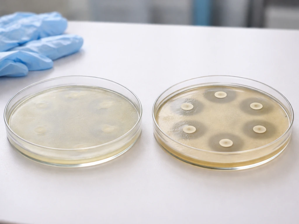
Yes, Staphylococcus aureus grows reliably on mannitol salt agar (MSA) and produces a distinctive yellow color change in the medium around its colonies. This is one of the classic results in introductory and applied microbiology: S. aureus tolerates the high salt concentration that kills most other organisms, and then ferments mannitol, driving down the pH and turning the phenol red indicator from red to bright yellow. If you're running an MSA plate and expecting that result, you should see yellow shiny colonies surrounded by a yellow zone after 24 to 48 hours at 35 to 37°C. Micrococcus luteus is not typically considered a typical grower on MSA in the way Staphylococcus aureus is.
Does S aureus Grow on MSA Results, Color, and Troubleshooting

Marcus Reeves
5 Jul 2026
What MSA is and how it works
Mannitol salt agar does two things at once: it selects for salt-tolerant organisms and differentiates them by whether or not they ferment mannitol. The selective part comes from a very high sodium chloride concentration, typically 75 g/L, which works out to about 7.5% NaCl. That's far above what most gram-negative bacteria and many gram-positive organisms can survive, so MSA acts as a gate that essentially only lets halotolerant (salt-tolerant) organisms through.
The differential part comes from two additional components: mannitol as a fermentable sugar, and phenol red as a pH indicator. The medium starts at a neutral pH of about 7.4, which keeps phenol red in its red/pink state. When an organism ferments mannitol, it produces acid, dropping the local pH and turning the phenol red indicator bright yellow. Organisms that grow but don't ferment mannitol leave the medium red or pink. This color shift is the key to interpreting your plate.
In practical terms, MSA is widely used in food safety labs, clinical microbiology, and teaching environments to screen for staphylococci, and particularly to get a presumptive signal for S. aureus. It's not a confirmatory test on its own, but it narrows things down quickly.
Does S. aureus grow on MSA? What to expect
S. aureus is well-adapted to high-salt environments, which is why MSA works so well as a presumptive screen. Most strains grow confidently at 7.5% NaCl without issue. More importantly, most S. aureus strains ferment mannitol, so you get both growth and a color change, which together form a presumptive positive result.
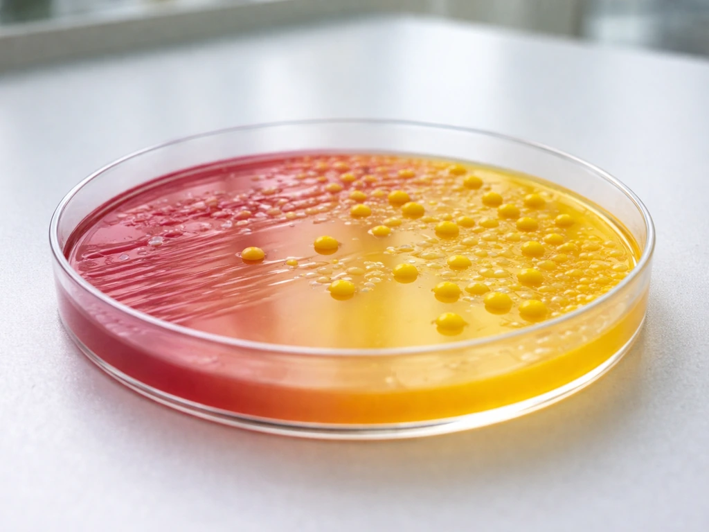
The quality control reference strain S. aureus ATCC 25923 is commonly used to validate MSA plates. On a properly prepared plate, it produces 1 to 2 mm yellow shiny colonies with a yellow zone spreading into the surrounding medium. The entire area around the growth should turn yellow, not just the colonies themselves. This is what a textbook positive looks like.
At standard incubation conditions (35 to 37°C, aerobic, 24 to 48 hours), the yellow color change is typically visible by 24 hours and fully developed by 48 hours. Some strains may take the full 48-hour window to show a strong result, so don't write off a plate at 24 hours if the yellowing looks weak or patchy.
Reading the plate: what the colors actually mean
There are essentially three outcomes when you read an MSA plate, and each tells you something specific.
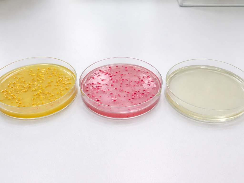
- Yellow colonies with a yellow zone in the surrounding medium: the organism grew (salt-tolerant) and fermented mannitol (acid production dropped the pH). This is a presumptive positive for S. aureus, though not definitive on its own.
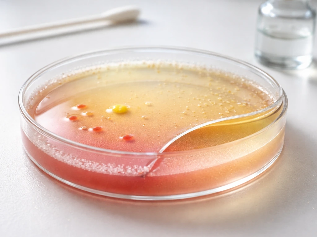
- Growth with a pink or red background and no yellow change: the organism is salt-tolerant but did not ferment mannitol. This pattern is typical of coagulase-negative staphylococci like S. epidermidis.
- No visible growth: the organism was inhibited by the high NaCl concentration, or the inoculum was too low, or conditions were not right for growth.
The yellow color change is caused by acid accumulating in the agar matrix around the colonies as mannitol fermentation proceeds. The phenol red indicator turns from red/neutral to yellow as pH drops. If you see partial or faint yellowing only directly under the colony, that can indicate slow or weak fermentation. Read the plate in good light and compare the medium color in growth zones against an uninoculated area of the plate to catch subtle shifts.
One thing worth keeping in mind: yellow on MSA means mannitol-fermenting and salt-tolerant, not automatically S. aureus. Other organisms with these traits can also produce yellow zones. S. aureus is the most common clinical and food-relevant cause of that result, but the color alone doesn't confirm species identity. Follow-up confirmation testing is essential.
Conditions that affect growth on MSA
Salt tolerance
S. aureus handles the 7.5% NaCl in MSA well because it's naturally halotolerant. This is part of what makes it such a relevant food safety pathogen: it can grow in cured meats, brined products, and other high-salt environments that would stop most pathogens in their tracks. On MSA, that tolerance is the gateway to growth.
Temperature
Incubate MSA plates at 35 to 37°C. Hardy Diagnostics specifies incubating mannitol salt agar aerobically at 35, 37°C for 24, 48 hours and notes that S. aureus appears as yellow colonies with yellow zones, while non-fermenters like S. epidermidis produce clear pink to red colonies with no yellow change incubate MSA aerobically at 35–37°C for 24–48 hours. This range supports both reliable growth and full mannitol fermentation. Some commercial protocols specify 35°C (such as CDC NHANES MRSA screening protocols), while others use 37°C. Both work. Going significantly below 35°C can slow fermentation enough that you won't see a clear color change within the normal reading window.
Incubation time
Standard incubation is 18 to 24 hours, with up to 48 hours recommended for full interpretation. Read the plate at 24 hours for an initial check, but hold the plate and re-read at 48 hours before calling a result negative or weakly positive. Some S. aureus strains show delayed mannitol fermentation, and a plate that looks borderline at 24 hours may be clearly yellow at 48 hours.
Oxygen availability
MSA plates should be incubated aerobically. Aerobic incubation is the standard condition specified across manufacturer IFUs and reference protocols. Staphylococci are facultative anaerobes, but fermentation performance and the color change reaction on MSA are optimized under aerobic conditions.
Why you might get a negative or unclear result
A few things can cause S. aureus to look negative or produce a confusing result on MSA. Most are fixable once you know what to look for.
- Reading too early: checking at 18 to 20 hours when the strain needs a full 48 hours. Always extend incubation before calling negative.
- Stressed or injured cells: organisms that have been heat-stressed, frozen, or exposed to sublethal sanitizers may have impaired growth or delayed fermentation. These injured cells can grow poorly on high-salt media even if they would normally thrive. Recovery broths or pre-enrichment steps help here.
- Inoculum too low: if you plate too few cells, colony numbers may be too sparse to generate enough acid to visibly shift the phenol red across the agar zone. A larger, well-distributed inoculum gives a cleaner result.
- Medium preparation errors: incorrect NaCl concentration, overheating the agar, or phenol red that has degraded can all cause failed selective or differential performance. A plate that doesn't turn yellow even with a confirmed S. aureus strain may have a medium preparation problem, not a growth problem.
- Rare mannitol-non-fermenting strains: not all S. aureus isolates ferment mannitol. A small proportion of strains are mannitol-use deficient and will grow on MSA (because they're still salt-tolerant) but will not produce yellow color change. This means no yellow result does not automatically rule out S. aureus.
- Contamination or competitive overgrowth: other halotolerant organisms in a mixed sample can outgrow S. aureus or produce their own color changes that obscure the result.
- Incubation temperature drift: incubating outside the 35 to 37°C range can slow fermentation and produce weak or absent color change even with viable organisms.
If your plate looks unusual, check your controls first. A positive control (S. aureus ATCC 25923) should show yellow colonies on every valid MSA run. If the control doesn't perform as expected, the problem is with your medium or conditions, not your sample.
How MSA results compare across Staphylococcus species
MSA was designed to differentiate S. aureus from coagulase-negative staphylococci (CoNS), and the color difference between them is usually clear. Here's how the most common Staphylococcus species typically behave.
| Organism | Growth on MSA | Mannitol Fermentation | Colony and Medium Appearance |
|---|---|---|---|
| S. aureus | Yes (good growth) | Yes (most strains) | 1–2 mm yellow shiny colonies, yellow zone in surrounding medium |
| S. epidermidis | Yes (may be slower) | No | ~1 mm white or pink shiny colonies, red/pink background, no yellow change |
| S. saprophyticus | Yes | Variable | Colonies present, medium color varies by strain |
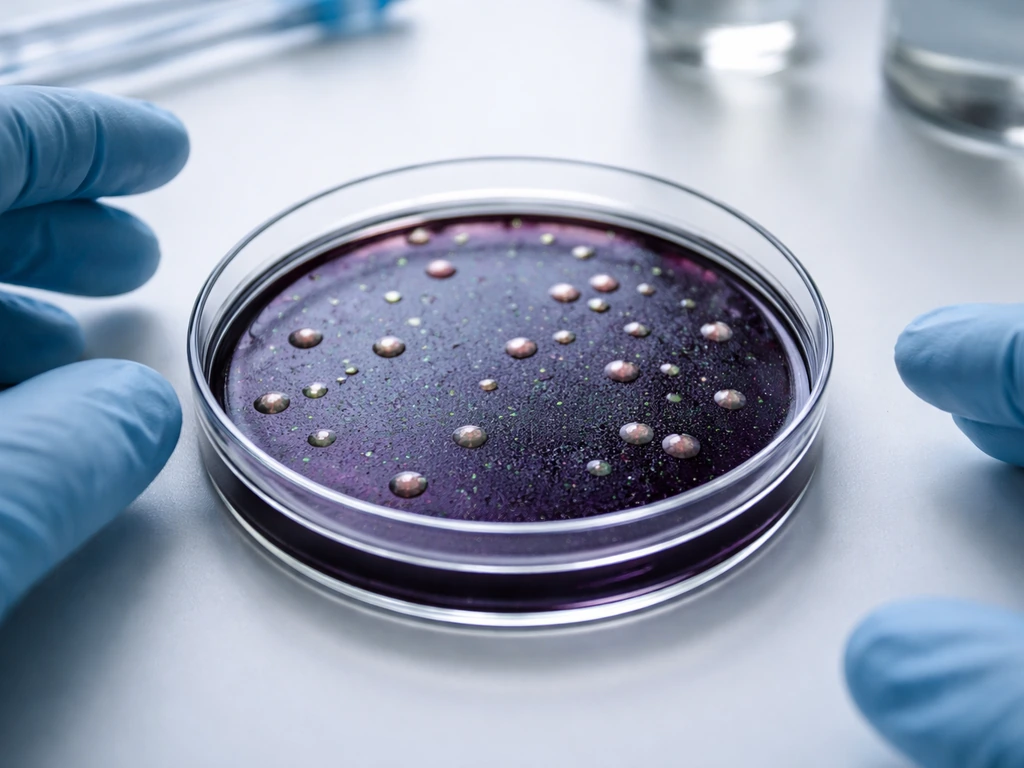
| Micrococcus luteus | Variable (some strains tolerate salt) | No | Colonies if present with red/pink background |
| Bacillus subtilis | Limited or partial inhibition | Variable | May show some growth; not a target organism for MSA |
| E. coli and most gram-negatives | Inhibited | N/A | No growth |
The most important distinction is between S. aureus and S. epidermidis. Both grow on MSA, but S. epidermidis does not ferment mannitol, so it leaves the medium red or pink. If you see growth but no yellow color change, you're likely looking at a non-pathogenic CoNS rather than S. aureus. That said, S. Bacillus subtilis can grow on mannitol salt agar (MSA), but you may not see the same reliable yellow color change you expect from S. aureus Bacillus subtilis grow on MSA. aureus and CoNS are not the only organisms that can survive on MSA. Other halotolerant bacteria, including some Micrococcus species, can also grow, which is one reason the medium is presumptive rather than confirmatory. The behavior of organisms like Micrococcus luteus on MSA is worth knowing if you're working with environmental or skin samples that carry mixed flora.
Next steps: confirming your MSA result
MSA is a presumptive tool. A yellow colony on MSA is strong evidence pointing toward S. aureus, but it's not a final identification. Unlike Staphylococcus, Serratia marcescens does not typically produce the mannitol-based yellow color change used to interpret MSA but it's not a final identification. The standard workflow is to use MSA as the first screen and then confirm with biochemical tests on isolated colonies.
- Pick yellow colonies from MSA and subculture to a non-selective agar (like tryptic soy agar or blood agar) to obtain a pure culture.
- Run a catalase test: S. aureus is catalase-positive (bubbles with hydrogen peroxide). This separates staphylococci from streptococci.
- Run a coagulase test: S. aureus is coagulase-positive (either tube coagulase or slide latex agglutination). This is the traditional gold-standard test for distinguishing S. aureus from CoNS.
- If MRSA is a concern, follow up with chromogenic MRSA agar or PCR-based confirmation depending on your setting and available resources.
- Always run a positive control (S. aureus ATCC 25923) and a negative control (S. epidermidis ATCC 12228) alongside your sample plates to validate each MSA run.
The controls are not optional if you're using results for any decision-making purpose. S. aureus ATCC 25923 should give you yellow colonies and a yellow medium zone. S. epidermidis ATCC 12228 should give you growth with a red or pink background and no color change. If either control behaves unexpectedly, recheck your medium preparation, incubation temperature, and phenol red functionality before trusting your sample results.
For food safety applications specifically, MSA is often used as part of a broader isolation workflow from food matrices. Pre-enrichment in a high-salt broth (such as 10% NaCl tryptic soy broth) before plating on MSA is common when working with processed foods, because injured cells in food matrices need recovery time before they can grow efficiently on selective media. Plating directly from a food homogenate without pre-enrichment can produce false negatives even when S. aureus is present.
Once you've confirmed S. aureus identity through coagulase testing, MSA growth behavior makes a lot more sense as one data point in a sequence rather than a standalone answer. The yellow color tells you something real and useful, but the complete picture always requires that follow-up step.
FAQ
How can I tell if a yellow zone on MSA is truly fermentation, not just a trick of the plate?
Look for yellowing spreading into the surrounding agar from the growth area, not only discoloration confined to the colony itself. Compare against an uninoculated area of the same plate under the same light conditions. If the whole growth zone is turning yellow but the colony sheen is faint, re-read at 48 hours and verify the phenol red indicator is functioning with your positive control.
If my plate has colonies that grow on MSA but no yellow color change, does that rule out S. aureus?
It strongly argues against S. aureus, but it does not fully rule it out. Some strains can ferment mannitol slowly, so a borderline result at 24 hours can become clearly yellow by 48 hours. Confirm identity with follow-up testing rather than relying on the initial color outcome alone.
What are the most common reasons S. aureus might appear negative on MSA?
The usual culprits are incubation conditions and medium quality. If plates are incubated below about 35°C, mannitol fermentation can be delayed and color may look weak by the expected reading time. Also check that the medium was prepared correctly and that phenol red is still active by running the required control strain each batch.
Does incubation atmosphere matter for the MSA mannitol color reaction?
Yes. MSA is typically incubated aerobically, and the mannitol fermentation and color development are most consistent under those standard conditions. While staphylococci can survive facultatively, deviating from aerobic conditions can lead to weaker or delayed color change.
Should I call an MSA plate negative if it looks red at 24 hours?
Not automatically. Many protocols recommend reading at about 24 hours as an initial check but holding up to 48 hours before calling it negative or weakly positive. If S. aureus is present with delayed fermentation, you can miss the yellow shift if you discard or report too early.
Can other bacteria besides S. aureus produce yellow colonies and a yellow zone on MSA?
Yes. Yellow on MSA indicates salt tolerance plus mannitol fermentation, not species identity. Coagulase-negative staphylococci are usually distinguished because they typically do not ferment mannitol, but other halotolerant organisms, including some Micrococcus species, can complicate interpretation. Use confirmation tests for final identification.
What is the best way to interpret mixed-flora samples where I see multiple colony morphologies?
Treat yellow zones as presumptive but evaluate each distinct colony type. When mixed colonies are present, record which colonies are associated with the yellow medium change, then isolate representative colonies for confirmation. Relying on the strongest-looking colony alone can mislead if another organism is driving the color change.
Do quality control strains need to match the exact incubation time window used for samples?
They need to be treated the same way as your test plates. If your workflow includes a 24-hour initial read and a 48-hour final read, run the controls through the same reading schedule. That way you can tell whether a weakly developing color pattern is expected for your conditions or indicates a problem with the medium or incubation.
Why is MSA considered presumptive instead of confirmatory for S. aureus?
Because the medium screens for salt tolerance and mannitol fermentation, which can overlap across different organisms and even within atypical strains. MSA is designed to narrow possibilities quickly, then coagulase testing and other confirmatory steps are used to reach species-level identification.
In food safety workflows, does pre-enrichment matter for getting an accurate MSA result for S. aureus?
It often matters. Injured or stressed cells from processed or high-salt environments may not grow well when plated directly, leading to false negatives. Pre-enrichment in a suitable high-salt broth before plating on MSA helps recovery and improves the chance of detecting S. aureus if it is present.
Next Article
Does Alcaligenes faecalis Grow on EMB Agar? Expected Results
Find out if Alcaligenes faecalis grows on EMB agar and what colony color patterns mean for ID troubleshooting.