
Yes, Listeria monocytogenes grows on chocolate agar. It is an enriched, non-selective medium, and L. monocytogenes has no trouble developing colonies on it under standard incubation conditions. Clinical laboratories routinely use chocolate agar plates to passage isolates, recover organisms from normally sterile specimens, and support confirmatory testing workflows. That said, chocolate agar has a significant practical limitation for Listeria work: because the red blood cells are lysed during preparation, you cannot observe the characteristic beta-hemolysis that helps identify and differentiate L. monocytogenes. For food safety and environmental testing, it also offers none of the selectivity or species-level differentiation that purpose-built Listeria media provide. So while the answer to the growth question is a straightforward yes, chocolate agar alone is rarely the right tool for detecting or confirming Listeria in a regulated testing context.
Does Listeria monocytogenes Grow on Chocolate Agar?

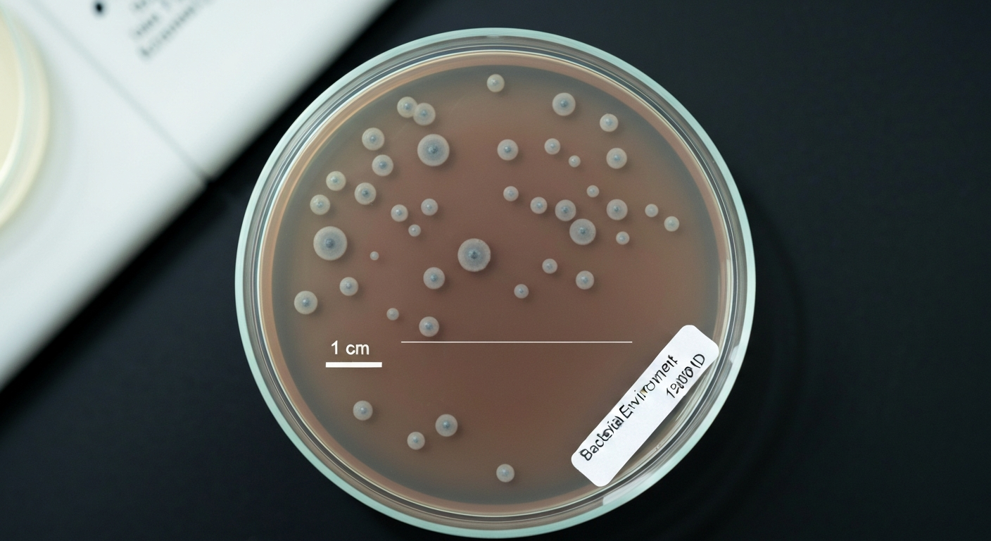
What chocolate agar is and how its composition supports bacterial growth
Chocolate agar is essentially heated blood agar. It is made by adding blood (typically sheep or horse) to a nutrient base and then raising the temperature to around 80 degrees Celsius. That heat lyses the red blood cells, releasing hemin (X factor) and NAD (V factor) into the medium and turning it the characteristic brown color that gives the agar its name. Chocolate agar is prepared by adding blood to a base medium and heating (lysing) the erythrocytes to release hemin (X factor) and NAD (V factor), i.e., chocolate agar = heated blood agar, which specifically supports fastidious organisms such as Neisseria and Haemophilus Chocolate agar is prepared by adding blood to a base medium and heating (lysing) the erythrocytes to release hemin (X factor) and NAD (V factor) — i.e., chocolate agar = heated blood agar — and is specifically formulated to support growth of fastidious organisms (Neisseria, Haemophilus).. The point of the lysis step is to make those growth factors freely available, which is why chocolate agar is the go-to medium for fastidious organisms like Neisseria gonorrhoeae and Haemophilus influenzae that cannot extract what they need from intact red cells.
For organisms that are not fastidious, like L. monocytogenes, the extra growth factors simply mean the medium is generous and forgiving rather than essential. Commercial formulations such as BD BBL Chocolate II also include hemoglobin and enrichment supplements like IsoVitaleX, which further broaden the range of organisms that will grow well. The key classification to keep in mind is that chocolate agar is an enriched, non-selective medium. It does not contain agents that suppress competing flora, so anything capable of growing on a standard nutrient agar will also grow here.
Conditions that allow L. monocytogenes to grow on chocolate agar
L. monocytogenes is psychrotrophic, meaning it can grow across a wide temperature range, from roughly 4 degrees Celsius up to about 45 degrees Celsius. Stress‑ and growth‑rate‑related differences between plate count and real‑time PCR data during growth of L. monocytogenes (AEM; growth kinetics) notes that incubation temperature strongly affects colony development speed, with growth possible at refrigeration (≈4 °C) and optimal growth near 30–37 °C. On chocolate agar, it will grow at standard clinical incubation temperatures of 35 plus or minus 2 degrees Celsius, which is where most routine isolate work takes place. Colony development is typically visible within 24 to 48 hours at that temperature. Enumeration workflows sometimes use 30 degrees Celsius instead, in line with ISO protocols, but for simple subculture and recovery purposes, 35 to 37 degrees Celsius is standard.
Chocolate agar is frequently incubated in a CO2-enriched atmosphere (around 5 percent CO2) when growing fastidious organisms, and this is fine for L. monocytogenes too, though it does not require CO2 to grow. Ambient air works. Most manufacturer instructions for chocolate agar products, including AccuProbe Listeria monocytogenes Culture Identification Test IFUs, explicitly list chocolate agar as an acceptable medium for growing Listeria colonies prior to testing, specifying 35 to 37 degrees Celsius incubation for 18 to 24 hours.
| Condition | Chocolate Agar Recommendation | Notes |
|---|---|---|
| Temperature | 35 ± 2 °C (routine); 30 °C (enumeration) | L. monocytogenes is psychrotrophic and will grow slowly even at 4 °C |
| Atmosphere | Ambient air or 5% CO2 | CO2 not required for Listeria; benefits fastidious co-isolates |
| Incubation time | 24–48 hours for visible colonies | Confirmatory reads typically at 48 h |
| Selectivity | None | All non-fastidious and fastidious organisms can grow freely |
What L. monocytogenes colonies look like on chocolate agar
On chocolate agar, L. monocytogenes produces small to medium, smooth, grayish-white to slightly translucent colonies after 24 to 48 hours at 35 degrees Celsius. The colonies are typically 1 to 2 mm in diameter and have a regular, convex shape with a moist, buttery appearance. Under oblique (Henry) illumination, which is a low-angle light technique used routinely in Listeria identification, the colonies show a bluish-green sheen that is a useful cue, though this is more dramatic on clear agars like tryptic soy agar.
The appearance on chocolate agar is not particularly distinctive compared to many other gram-positive rods. Without hemolysis being visible and without selective suppression of competing organisms, you cannot make a presumptive identification from colony morphology alone on this medium. It tells you something grew, but not what.
Why hemolysis disappears on chocolate agar and why that matters
This is the central interpretive problem with using chocolate agar for Listeria work. Beta-hemolysis, the clear zone of complete red cell lysis that surrounds L. monocytogenes colonies on blood agar, is produced by listeriolysin O (LLO), a cholesterol-dependent cytolysin encoded by the hly gene. On sheep blood agar, this zone is typically narrow and close to the colony, but it is visible and diagnostically meaningful. On chocolate agar, the red cells have already been lysed during media preparation, so there are no intact cells left to lyse. The hemolytic activity of the organism simply goes undetected.
This matters because hemolysis is one of the primary phenotypic features used to differentiate L. monocytogenes (beta-hemolytic) from non-hemolytic Listeria species like L. innocua, which can otherwise produce similar colony morphology. It also informs the CAMP test, which exploits the interaction between LLO and Staphylococcus aureus or Rhodococcus equi to produce enhanced hemolysis zones. None of this is visible on chocolate agar. Any laboratory workflow that needs to confirm L. monocytogenes must transfer candidate colonies to 5 percent sheep blood agar for hemolysis assessment.
It is also worth noting that hemolytic expression is strain-dependent. Inter-strain variability in LLO secretion means some L. monocytogenes isolates produce only a weak or narrow zone even on sheep blood agar. This variability reinforces why standardized confirmatory tests and selective media are preferred over relying on hemolysis observation alone, whether on blood agar or elsewhere.
Blood agar vs. chocolate agar: what each one actually gives you
Blood agar (5 percent sheep blood agar) and chocolate agar share a common starting point but serve different diagnostic purposes once the heating step is introduced or omitted. On blood agar, the red cells are intact, hemolysis is visible, and L. monocytogenes produces its characteristic narrow beta-hemolytic zone, which you read by direct examination and by the stab method (pushing a loop vertically into the agar surface to demonstrate subsurface hemolysis). Blood agar is therefore the correct medium for hemolysis confirmation and the CAMP test.
A blood agar formulation called Listeria monocytogenes blood agar (LMBA) has been used specifically to exploit this hemolytic differentiation. A comparative study published in Food Control (2001) found that LMBA recovered more L. monocytogenes positives from naturally contaminated food and environmental samples than either PALCAM or Oxford agar in some sample sets, suggesting that blood-based hemolysis expression can sometimes capture strains that selective agars miss. This does not mean blood agar should replace selective media, but it does illustrate that hemolysis visibility has real detection value.
Chocolate agar, by contrast, is better suited for primary enriched culture from sterile clinical specimens (where competing flora is not a concern) or for subculturing and passaging isolates that have already been identified. It supports growth robustly and is comfortable for the organism, but it contributes nothing to identification beyond confirming that something grew.
| Feature | Blood Agar (5% Sheep) | Chocolate Agar |
|---|---|---|
| Red blood cells | Intact | Lysed (heated) |
| Beta-hemolysis visible | Yes | No |
| CAMP test compatible | Yes | No |
| Selectivity | None | None |
| Best use for Listeria | Hemolysis confirmation, CAMP test, subculture | Primary enriched culture, isolate passage, AccuProbe-type testing |
| Supports fastidious organisms | Partially | Yes (X and V factors released) |
How selective and chromogenic agars compare for Listeria detection
For food safety and environmental testing, the agars that actually do the heavy lifting are esculin-based selective media (Oxford and PALCAM) and chromogenic Listeria differential agars (ALOA, CHROMagar Listeria, R&F LMCPM, and similar). These are purpose-designed for Listeria and offer a combination of selectivity and species-level differentiation that neither blood agar nor chocolate agar can match.
Oxford and PALCAM agars
Both Oxford and PALCAM agars contain esculin, which Listeria spp. hydrolyze to produce a black precipitate (esculin + ferric ions), giving Listeria colonies a characteristic black halo. Oxford agar uses a combination of selective antibiotics (acriflavine, cefotetan, colistin, fosfomycin, and others) to suppress competing gram-negative and gram-positive organisms. PALCAM uses a different antibiotic mix and includes mannitol with phenol red as an indicator. On both media, Listeria colonies typically appear gray-green to olive with a black halo. These media distinguish Listeria at the genus level but do not reliably differentiate L. monocytogenes from other Listeria species by colony appearance alone.
Chromogenic Listeria agars (ALOA and equivalents)
Chromogenic media like ALOA (Agar Listeria Ottaviani Agosti) and CHROMagar Listeria detect phosphatidylinositol-specific phospholipase C (PI-PLC) activity, which is expressed by L. monocytogenes and L. ivanovii but not most other Listeria species. PI-PLC cleaves a chromogenic substrate in the medium, producing a blue or blue-green colony with an opaque white halo around it. This halo is a strong presumptive indicator for L. monocytogenes specifically. Validation studies have shown that chromogenic media improve speed and ease of presumptive species-level identification compared to esculin-based media alone.
One important caveat: multiple collaborative studies have documented that some L. monocytogenes strains, particularly stressed, injured, or atypical isolates, give weak or absent reactions on selective and chromogenic media. They may produce atypical colony morphology, faint halos, or be outcompeted even on selective plates. This is why the standard practice is to plate onto both an esculin-based medium and a chromogenic medium simultaneously, and why confirmation steps cannot be skipped.
| Medium | Type | L. monocytogenes Colony Appearance | Species Differentiation | Hemolysis Visible |
|---|---|---|---|---|
| Chocolate agar | Enriched, non-selective | Small, gray-white, smooth | No | No |
| Blood agar (5% sheep) | Enriched, non-selective | Small, gray-white with narrow beta-hemolytic zone | Partial (via hemolysis/CAMP) | Yes |
| Oxford agar | Selective, esculin-based | Gray-green with black halo | Genus level only | No |
| PALCAM agar | Selective, esculin-based | Olive/gray with black halo | Genus level only | No |
| ALOA / chromogenic Listeria | Selective + chromogenic | Blue-green with opaque white halo | Presumptive L. monocytogenes vs. other Listeria | No |
A note on related organisms and how they behave on chocolate agar
Because chocolate agar is a general enriched medium, many other organisms relevant to food safety and clinical microbiology also grow on it. Pseudomonas aeruginosa also grows readily on enriched media; see how does pseudomonas aeruginosa grow for growth characteristics and culture conditions. E. coli grows readily on chocolate agar, producing characteristic colonies, though just as with Listeria, there is no selectivity to aid identification. For more detail on E. coli growth on chocolate agar, see does e coli grow on chocolate agar. Pseudomonas aeruginosa grows well on chocolate agar and may produce its characteristic pigmentation under appropriate conditions, though it is typically identified on more specific media or by biochemical profiling. Group B Streptococcus (Streptococcus agalactiae) also grows on chocolate agar, but like L. monocytogenes, its hemolytic characteristic cannot be assessed there, requiring transfer to blood agar for confirmation. In all of these cases, chocolate agar supports growth without providing identification, which is the consistent limitation of the medium across organism types.
Where chocolate agar fits in Listeria detection workflows
Regulated testing workflows for L. monocytogenes in food and environmental samples are defined by FDA BAM Chapter 10 and ISO 11290. Both prescribe a multi-step process that begins with enrichment, moves to selective plating on two different agar types, and then requires confirmatory testing of presumptive colonies. Chocolate agar does not appear in these workflows as a primary plating medium. Here is how the process works in practice.
Step 1: Enrichment
Food and environmental samples typically carry low numbers of Listeria cells, often in the presence of large numbers of competing organisms. Direct plating without enrichment routinely fails to detect the target. FDA BAM uses a two-stage enrichment: an initial growth in University of Vermont Medium (UVM) or Buffered Listeria Enrichment Broth (BLEB), followed by secondary enrichment in Fraser broth. ISO 11290 uses half-Fraser broth for primary enrichment and full Fraser broth for secondary enrichment. Both systems use selective agents (nalidixic acid, acriflavine, and others in FDA BAM formulations; the same class in ISO formulations) that suppress background flora while allowing Listeria to multiply to detectable levels.
Step 2: Selective plating
After enrichment, cultures are plated onto at least one esculin-based selective agar (Oxford or PALCAM) and one chromogenic Listeria agar (ALOA or an approved equivalent). Plates are incubated at 35 to 37 degrees Celsius for 24 to 48 hours. Plating onto both agar types simultaneously is specified because each medium has documented sensitivity and specificity tradeoffs, and using both increases the probability of recovering atypical or stressed strains that might be missed on one medium alone.
Step 3: Confirmation
Presumptive Listeria colonies from the selective plates are picked and streaked onto 5 percent sheep blood agar for hemolysis assessment. The stab method is used to demonstrate subsurface beta-hemolysis, and the CAMP test is run against S. aureus and R. equi to confirm L. monocytogenes specifically. Additional confirmatory tests include carbohydrate fermentation (xylose negative, rhamnose positive for L. monocytogenes), motility at 25 degrees Celsius, gram stain, and catalase test. Molecular confirmation by PCR targeting the hly gene (LLO) or other specific loci is increasingly routine.
Chocolate agar can have a role at the confirmation stage, specifically for subculturing isolates, performing AccuProbe or similar culture identification tests, or passaging confirmed colonies. It is a reliable growth medium at that point, and manufacturer IFUs for several diagnostic kits explicitly list chocolate agar as an approved substrate for growing colonies before testing. What it cannot do is substitute for selective plating, enrichment, or hemolysis-based confirmation in any step of the regulated workflow.
The full workflow at a glance
- Primary enrichment: UVM/BLEB (FDA BAM) or half-Fraser broth (ISO 11290), 24–26 hours at 30 °C
- Secondary enrichment: Fraser broth (FDA BAM) or full Fraser broth (ISO 11290), 48 hours at 37 °C
- Selective plating: Oxford or PALCAM agar AND ALOA or approved chromogenic Listeria agar, 24–48 hours at 35–37 °C
- Presumptive identification: Read esculin hydrolysis (black halo) and PI-PLC chromogenic reaction (blue-green colony with opaque halo on ALOA)
- Confirmation on blood agar: Stab test for beta-hemolysis, CAMP test on 5% sheep blood agar
- Biochemical confirmation: Carbohydrate fermentation, motility, catalase, gram stain
- Molecular confirmation (where required): PCR for hly or species-specific targets
- Optional: Subculture on chocolate agar for diagnostic kit use (AccuProbe, etc.) or isolate passage
Practical takeaways for the lab
If you are working with a confirmed L. monocytogenes isolate and need a reliable general-purpose plate for subculture or downstream testing, chocolate agar is a solid choice. It supports robust growth at 35 degrees Celsius within 24 to 48 hours, and several molecular and culture-identification kits approve it specifically. If you are trying to detect, screen, or confirm Listeria from food, environmental swabs, or clinical samples where you do not already know the isolate identity, chocolate agar by itself will not get you there. You need enrichment, selective plating on esculin and chromogenic Listeria media, and confirmatory hemolysis work on sheep blood agar. Chocolate agar fits into that workflow as a supporting medium, not a substitute for any of the required steps.
FAQ
Does Listeria monocytogenes grow on chocolate agar?
Yes. Listeria monocytogenes will grow on chocolate agar (heated/lysed blood agar). Chocolate agar is an enriched, nonselective medium that supports growth of many bacteria including L. monocytogenes.
Under what environmental and cultural conditions will L. monocytogenes grow on chocolate agar?
L. monocytogenes grows on chocolate agar across the organism's normal laboratory growth range. Typical incubation conditions used in clinical labs are 35±2 °C for 24–48 h in ambient air (CO2 is not required). The organism is psychrotrophic (can grow at refrigeration temperatures), but colony development on plates is much slower at 4–8 °C. Chocolate agar formulations are enriched (lysed blood/hemin/NAD supplements) and provide sufficient nutrients for growth; incubation time and temperature determine how fast colonies become visible.
What colony morphology should I expect for L. monocytogenes on chocolate agar?
Colonies are typically small to medium, gray-white, smooth and translucent to slightly opaque. Colony size and appearance vary by strain and incubation time; growth may be slower for stressed or injured cells. Chocolate agar will not produce the characteristic hemolytic clearing because red cells are lysed during manufacture.
How does chocolate agar affect observation of hemolysis for L. monocytogenes?
Hemolysis cannot be visually assessed on chocolate agar because the erythrocytes have been lysed when the medium was prepared. Listeriolysin O–mediated β‑hemolysis is therefore not visible on chocolate plates; hemolysis (a narrow β‑hemolytic zone) must be evaluated by subculture onto 5% sheep blood agar (streak and stab methods) or by using specific hemolysis‑revealing techniques.
How does chocolate agar compare to blood agar and Listeria‑selective agars for detecting L. monocytogenes?
Chocolate agar is an enriched, nonselective medium that will grow L. monocytogenes but does not allow hemolysis assessment. 5% sheep blood agar shows β‑hemolysis useful for presumptive ID. Selective Listeria media (Oxford, PALCAM) and chromogenic Listeria agars (ALOA/CHROMagar Listeria) contain selective agents and differential substrates that increase specificity and help suppress background flora. Regulatory and reference methods use enrichment plus plating onto esculin‑based selective agar and chromogenic Listeria agar because these media increase recovery and presumptive identification relative to nonselective agars alone.
What are the recommended workflows for detection of L. monocytogenes in food and environmental testing?
Follow validated/reference workflows: perform primary enrichment (e.g., half‑Fraser/Fraser or FDA/BAM enrichment schemes), then plate onto at least one esculin‑based selective agar (Oxford or PALCAM) and one chromogenic Listeria differential agar (e.g., ALOA). Incubate per method instructions and pick presumptive colonies for confirmatory testing (subculture to 5% sheep blood agar to check hemolysis, biochemical tests, serology, and/or PCR). Using both selective and chromogenic plates reduces false negatives from selective‑media variability.
How Does Pseudomonas aeruginosa Grow? Conditions to Know
Learn how Pseudomonas aeruginosa grows and persists: temperature, pH, moisture, oxygen, nutrients, biofilms, and prevent